22 prasad etal
TRANSCRIPT
7/27/2019 22 Prasad Etal
http://slidepdf.com/reader/full/22-prasad-etal 1/6
897
Prasad et al., Int J Med Res Health Sci. 2014;3(4):897-902
International Journal of Medical Research
&
Health Sciences
www.ijmrhs.com Volume 3 Issue 4 Coden: IJMRHS Copyright @2014 ISSN: 2319-5886Received: 16
thAug 2014 Revised: 18
thSep 2014 Accepted: 29
thSep 2014
Research article
AN INEXPENSIVE AND INNOVATIVE CORRECTION OF MEDIAL COMPARTMENTAL
OSTEOARTHRITIS KNEE JOINT BY HIGH TIBIAL LATERAL CLOSED WEDGE OSTEOTOMY IN
A RURAL SET UP
Prasad DV1,*Arun AA
2, Tushar Chaudhari
2, Sagar Jawale
2, Shakthi Panda
2, Abhinav Jadhav
2, Deepak Dathrange
2
1Professor,
2Resident, Dept of Orthopaedics, Rural Medical College and Pravara Rural Hospital, Loni,
Maharashtra, India
*Corresponding author email: [email protected]
ABSTRACT
Osteoarthritis of Knee joint with Varus deformity causes considerable disability. Operative treatment aims at
shifting the mechanical load bearing axis to the less affected compartment of the knee to relieve the symptoms.
Exclusion Criteria: Non-walkers due to generalized arthropathies / medical comorbidities, Flexion deformity > 10
degrees, Range of motion < 90 degrees, Active rheumatoid arthritis, Infection, Lateral compartment involvement,
>1cm lateral subluxation in standing A-P X rays of both knees. Methodology: 32 (12 Males and 20 Females)
cases of Medial compartment osteoarthritis presenting in our OPD between 2008-2012 were treated by HTOand
cortical screw and SS wire fixation (TBW Technique). Results: Evaluation of results was done based on knee
rating scale by Japanese orthopaedic association. 22 cases were Excellent, 8 cases were good. One case of failure,
an iatrogenic intracondylar fracture of Tibia, and another secondary haematoma under the suture line, aspirated
and complete healing was achieved. Patients had good range of motion, were able to squat and sit cross legged
comfortably. Conclusion: HTO by Closed Medial wedge osteotomy and fixation with cortical screw and SS wire
provides a good alternative to unicompartmental knee Arthroplasty and even Total knee Arthroplasty (may be up
to 10-15 years) in patients with Medial compartmental osteoarthritis. It is a cost effective technique with the use
of minimum hardware and early postoperative mobilization in patients who cannot afford Knee Arthroplasty in a
Rural set up.
Keywords: Medial Compartmental Osteoarthritis, High Tibial osteotomy (HTO), Tension Band Wiring (TBW).
INTRODUCTION
Osteoarthritis of the Knee is a Chronic debilitating
disease excessive pressure leads to breakdown of the
cartilage matrix, architectural changes in the
subchondral bone, further altering the joint
geometry1,2
. Most of the patients present with
unicompartmental osteoarthritis (Medial
compartment) with varus deformity compromising
their day to day activities and finally leading topainful arthrosis. Prevalence of osteoarthritis of knee
is 5% to 13% in India. Our cultural and religious
habits and daily activities need most of the Indians,
particularly in the rural side to squat and sit cross
legged. With unicompartmental or total arthroplasty
sitting cross legged or squatting are
restricted3.Osteotomy of the tibia was originally used
to address osteoarthritis of the knee with an objective
to shift load bearing from one arthritic tibiofemoral
compartment to the other less affected compartment4,
5,6(Unloading of Involved Joint) Whereas in HTO
(using double TBW and 2 cortical screws with
DOI: 10.5958/2319-5886.2014.00022.8
7/27/2019 22 Prasad Etal
http://slidepdf.com/reader/full/22-prasad-etal 2/6
898
Prasad et al., Int J Med Res Health Sci. 2014;3(4):897-902
washers), patient is mobilised in immediate
postoperative period and patient can resume sitting
cross legged and squatting. This procedure is cheap
and cost effective as compared to other procedures,
the cost of implants being approximately Rs.500 to
600 only. One of the biggest advantages of HTO is
need for knee arthroplasty can be postponed for a
minimum period of 12- 15 years as found in
literature.3
The knee consists of 3 compartments: the
medial tibio-femoral, the lateral tibio-femoral and
patello-femoral. Out of which medial tibio-femoral
compartment is most commonly affected1. We
studied the effect of TBW with 2 cortical screws in
32 cases of uni-compartmental O.A. of Knee joint.
Biomechanics: In the standing position and chiefly
during walking, the body weight tends to adduct the
femur on the tibia, increasing thus the load on the
medial compartment. The lateral muscular forces tend
to adjust a dynamic equilibrium in the knees. The
lateral force and the body weight result in an overload
distribution of about 60% in the medial compartment
and 40% in the lateral compartment. In medial
compartment arthritis; the resulting lateral force is
displaced medially. Limb alignment is altered and
more loads are then distributed medially with
subsequent degenerative lesions. This progressive
joint destruction causes knee deformity1,7, which, in a
vicious circle, aggravates arthritis in the medial
compartment, resulting in a varus deformity at knee
joint. Various methods of treatment in High Tibial
Osteotomy (HTO) are- plaster cast, External fixator,
Coventry plate, Broad dynamic compression plate,
Locking compression plate, Joshi's external
stabilization system, TBW (minimal
instrumentation)8. Jackson was the first to report his
experience with femoral and tibial osteotomies to
treat osteoarthritis with associated valgus and varusknee alignment
8,9. Tension Band Wiring Principle in
HTO: By using double TBW distracting forces are
converted into converging forces at the lateral closed
wedge osteotomy site by anchoring the Tension band
around the Iliotibial band at its attachment at the
Gerdy’s tubercle which helps in early mobilization of
the patient.
MATERIALS AND METHODS
32 cases (12 Males and 20 Females) of Medialcompartment osteoarthritis, majority between the age
group of 50-65 years presenting in the OPD of Rural
Medical college and hospital, Loni between the
period of 2008-2013, were treated by High Tibial
closed wedge osteotomy and 2 cortical screw, 2
washers and Double Stainless Steel wire fixation
(TBW Technique). Japanese Orthopaedic association
scoring was used for assessment of cases after taking
ethical clearance.
Inclusion criteria: People with degenerative disease
of knee (osteoarthritis) between the age of 45 to 65
years. a) Who are walking independently or with one
stick. b) Who accepted “repairing” the joint than
“replacing”. Male and Female patients between 55-70
years age, having Medial compartmental
osteoarthritis presenting with Pain. Patients with
Knee Flexion up to 90° with full extension possible.
Patients able to squat and sit cross legged. A written
informed consent of the participants was taken before
initiation of the study.
Exclusion criteria: Non walkers due to generalised
arthropathies or medical morbidity. Flexion deformity
of knee more than 10 degrees or range of motion less
than 90 degrees, Active rheumatoid arthritis or active
infection, Grossly symptomatic lateral compartment
involvement, more than 1 cm lateral subluxation of
tibia as judged by standing AP x-rays of both knees.
Grossly advanced arthritis or tricompartment arthritis.
Preoperative Assessments
A) Preoperative Planning
i. Patient assessment: Patient’s age, career, level of
activity, previous history of surgery on the knee, and
expectation were taken into consideration before
deciding upon surgery. Closing wedge HTO may be
more beneficial in reducing the risk of nonunion than
opening wedge HTO for heavy-smoking patients10.
The ROM, degree of deformity, ligamentous
instability, and leg length discrepancy should be
assessed through physical examination. Valgus HTOcan be performed for minor or moderate medial
instability that can be caused by bone loss in medial
compartment osteoarthritis. The status of the hip joint
can have an influence on the medial osteoarthritis of
the ipsilateral knee. Abduction of the hip that occurs
during the stance period increases stress on the lateral
compartment of the knee, which gives rise to the
involvement of the stabilizers (gluteus maximus,
tensor fascia latae, and biceps femoris) that results in
higher forces on the lateral knee
11.
Therefore, hipabductor muscle weakness or restriction or ankylosis
of the hip joint should be treated prior to HTO.
7/27/2019 22 Prasad Etal
http://slidepdf.com/reader/full/22-prasad-etal 3/6
899
Prasad et al., Int J Med Res Health Sci. 2014;3(4):897-902
ii. Radiographic assessment: Multiple views should
be obtained for preoperative radiographic assessment:
bilateral weight-bearing anterior-posterior views in
full extension, tunnel views with the knee in 30o
of
flexion, Rosenberg views with the knee in 45o
of
flexion, lateral views, and skyline views. The severity
of medial osteoarthritis and bone loss evaluated from
the anterior-posterior views and patellar height
measured from the lateral views. A severe patella alta
may necessitate the combined use of tibial tubercle
osteotomy and closing/opening HTO. Lower limb
alignment is accessed from the full length
radiographs of the lower extremity that visualizes the
alignment of the hip, knee, and ankle joints. Magnetic
resonance imaging can be helpful in detecting
intraosseous lesions, meniscal tears, ligamentous
lesions, osteochondral defects, osteonecrosis, or
subchondraledema.
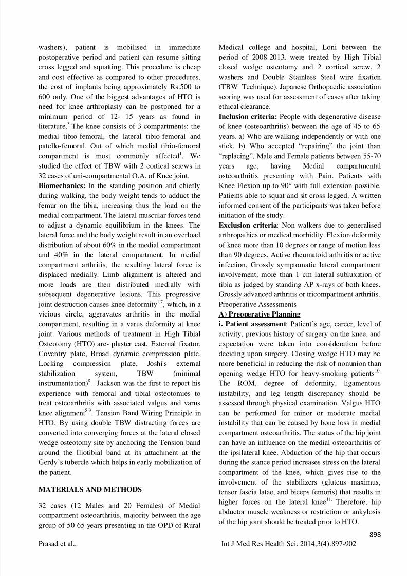
Fig 1: Showing the mechanical and anatomical axis
iii. Correction angle calculation: In normal lower
extremities, the centre of the hip is in line with the
centre of the knee and the centre of the ankle and the
mechanical axis, a line that connects the dots, is 0o
(Fig. 1). The ideal postoperative lower limb
alignment is considered as 3o-5
oof valgus from the
mechanical axis or 8o-10
oof anatomical valgus in
most studies. The Correction angle (α) is calculated in
the standing position as postulated by Fujisawa Y,
Masuhara K, ShiomiS12
or in the supine position as
postulated by K. Ogata, I. Yoshii, H. Kawamura et al.
The distal osteotomy line is determined referring to
the α angle and the wedge bone between the
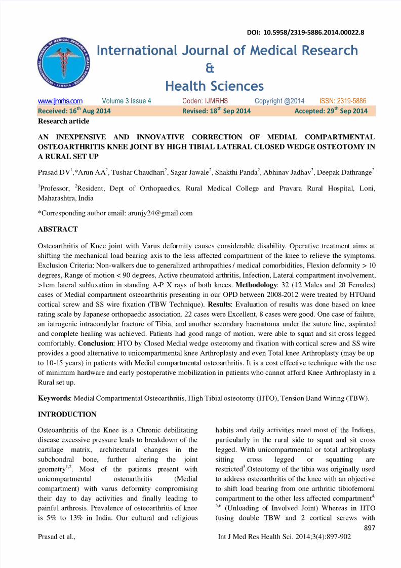
osteotomy lines is removed (Fig. 2A).
Fig 2A: showing calculation of α angle &2B showing
the endpoints used for exposure during surgery
Opening wedge HTO is planned in a similar fashion
like the closing wedge HTO. The proximal osteotomy
line is drawn from a point 3-4.5 cm inferior to themedial knee joint line to the tip of the fibular head
from which another same length line is drawn
obliquely by the α angle. The line that runs between
the endpoints of each line is used for exposure during
surgery (Fig. 2a, 2B). In HTO for medial
compartment osteoarthritis without knee instability,
efforts should be made to maintain the preoperative
anatomical posterior tibial slope. Some recent clinical
studies have shown that the use of navigation systems
contributed to the preciseness, accuracy, and
reproducibility of HTO.
B) HTO Techniques: There are various HTO
techniques including closing wedge osteotomy,
opening wedge osteotomy, dome osteotomy,
progressive callus distraction, and chevron
osteotomy. Of these, opening wedge HTO and
closing wedge HTO are most commonly performed
Medial Opening Wedge Osteotomy13
Surgical technique: The patient is placed in the
supine position on a radiolucent operating table and a
tourniquet is applied. A 5-cm vertical incision is
made over the center between the medial aspect of
the tibial tuberosity and the posteromedial aspect of
the tibia below the joint line. The pesanserinus is
detached from the tibia to expose the superficial
medial collateral ligament. The distal portion of the
exposed ligament is separated from bone and a blunt
retractor is inserted posterior to the medial collateral
ligament and the tibia to protect the neurovascular
structures posterior to the incision line. After
identifying the medial border of the patellar tendon,subperiosteal dissection is performed from the tibial
tuberosity to the posteromedial aspect of the tibia.
7/27/2019 22 Prasad Etal
http://slidepdf.com/reader/full/22-prasad-etal 4/6
900
Prasad et al., Int J Med Res Health Sci. 2014;3(4):897-902
Two guide wires are inserted at a point 3.5-4 cm
below the medial joint line and passed obliquely 1 cm
below the lateral articular margin of the tibia towards
the tip of the fibular head. After checking the
appropriate location with a fluoroscope, a tibial
osteotomy is performed immediately below the guide
wires using an oscillating saw or an osteotome.
Ensure the osteotomy line extends from the tibial
tuberosity along the posteromedial aspect of the tibia
to 1 cm medial to the the lateral tibial cortex and is in
parallel with the posterior tibial slope on the sagittal
plane. The mobility of the osteotomy site is checked
and the osteotomy is opened with a valgus force. If
the opening of the osteotomy seems insufficient, use
2 or 3 stacked osteotomes to reduce the risk of
intraarticular fractures. Subsequently, a calibrated
wedge is inserted until the osteotomy is opened to the
desired extent. Ensure with fluoroscopy when a long
alignment rod or wire cable is cantered over the hip
joint and the ankle joint, it lies at 62.5% of the width
of the tibial plateau. Once the desired degree of
correction is achieved, internal fixation of a metal
plate is performed. There are various types of metal
plates, including the Puddu plate, Tomofix, Aesculap
(dual) plate, -plates with or without a spacer
(rectangular or tapered). Among these, spacer plates
are most commonly used and the metal block should
be identical to the calibrated wedge. The proximal
fixation screws should be used under fluoroscopic
guidance and the defect should be grafted using iliac
crest autograft, allograft, or a bone substitute. For
defects ≥10 mm, cortico cancellous autografts or
allografts are used, whereas for small defects, bone
grafting is optional.
Other Techniqu: : Other HTO techniques include
dome osteotomy, progressive callus distraction using
an external fixator, and chevron osteotomy
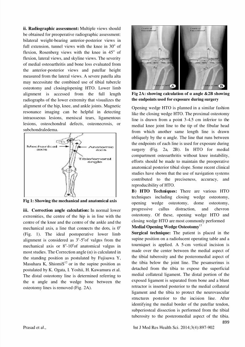
Fig 3: Showing the pre operative clinical and x-ray images
Our Operative Technique: After assessing the patient
clinically and radiographically as mentioned above
the patient is posted for corrective surgery.
In Supine position, on a radiolucent table under
image intensifier the proximal aspect of tibia was
approached through Lateral inverted L shapedincision. Close lateral wedge osteotomy was done and
angle of the wedge to be removed was determined pre
operatively with the help of radiographs. Lateral
wedge osteotomy done 1.5 cm distal to joint margin
to avoid fracture of tibial plateau intraoperatively
(Fig.4). Height of wedge is taken dependent on varus
angle calculated from x-rays, for each degree 1 mm
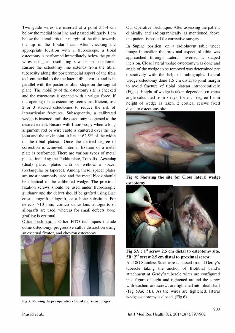
height of wedge is taken. 2 cortical screws fixed
distal to osteotomy site.
Fig 4: Showing the site for Close lateral wedge
osteotomy
Fig 5A : 1st
screw 2.5 cm distal to osteotomy site.
5B: 2nd
screw 2.5 cm distal to proximal screw.
An 18G Stainless Steel wire is passed around Gerdy’s
tubercle taking the anchor of Iliotibial band’s
attachment at Gerdy’s tubercle wires are configured
in a figure of eight and tightened around the screw
with washers and screws are tightened into tibial shaft
(Fig 5A& 5B). As the wires are tightened, lateralwedge osteotomy is closed. (Fig 6)
7/27/2019 22 Prasad Etal
http://slidepdf.com/reader/full/22-prasad-etal 5/6
901
Prasad et al., Int J Med Res Health Sci. 2014;3(4):897-902
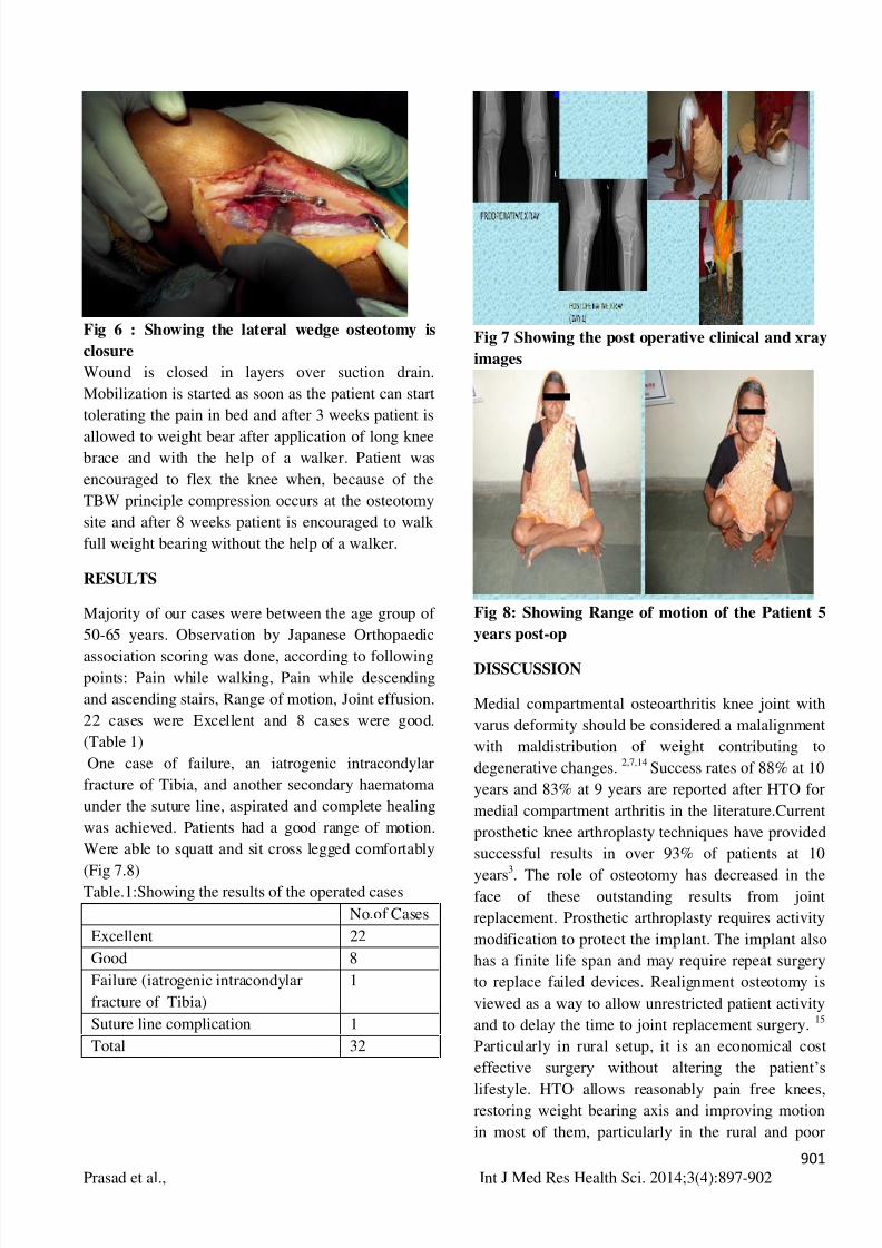
Fig 6 : Showing the lateral wedge osteotomy is
closure
Wound is closed in layers over suction drain.
Mobilization is started as soon as the patient can start
tolerating the pain in bed and after 3 weeks patient is
allowed to weight bear after application of long kneebrace and with the help of a walker. Patient was
encouraged to flex the knee when, because of the
TBW principle compression occurs at the osteotomy
site and after 8 weeks patient is encouraged to walk
full weight bearing without the help of a walker.
RESULTS
Majority of our cases were between the age group of
50-65 years. Observation by Japanese Orthopaedic
association scoring was done, according to followingpoints: Pain while walking, Pain while descending
and ascending stairs, Range of motion, Joint effusion.
22 cases were Excellent and 8 cases were good.
(Table 1)
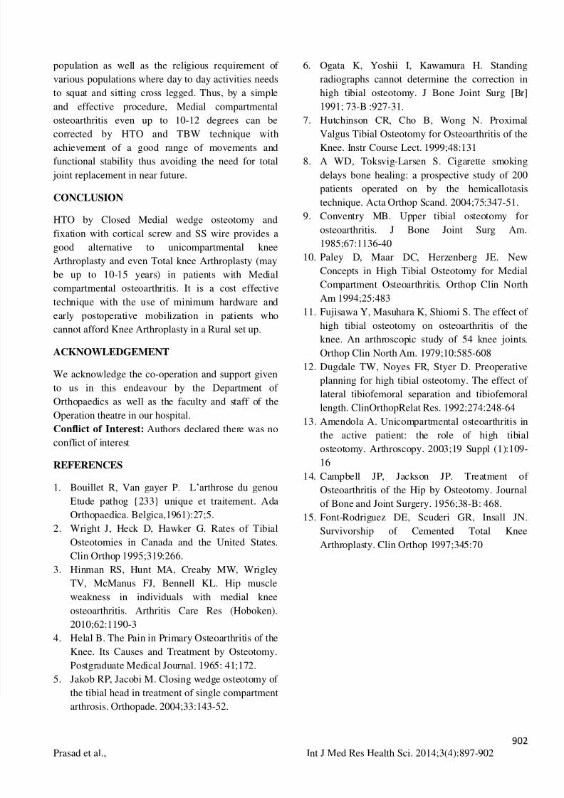
One case of failure, an iatrogenic intracondylar
fracture of Tibia, and another secondary haematoma
under the suture line, aspirated and complete healing
was achieved. Patients had a good range of motion.
Were able to squatt and sit cross legged comfortably
(Fig 7.8)Table.1:Showing the results of the operated cases
No.of Cases
Excellent 22
Good 8
Failure (iatrogenic intracondylar
fracture of Tibia)
1
Suture line complication 1
Total 32
Fig 7 Showing the post operative clinical and xray
images
Fig 8: Showing Range of motion of the Patient 5
years post-op
DISSCUSSION
Medial compartmental osteoarthritis knee joint with
varus deformity should be considered a malalignment
with maldistribution of weight contributing to
degenerative changes.2,7,14
Success rates of 88% at 10
years and 83% at 9 years are reported after HTO for
medial compartment arthritis in the literature.Current
prosthetic knee arthroplasty techniques have provided
successful results in over 93% of patients at 10
years3
. The role of osteotomy has decreased in theface of these outstanding results from joint
replacement. Prosthetic arthroplasty requires activity
modification to protect the implant. The implant also
has a finite life span and may require repeat surgery
to replace failed devices. Realignment osteotomy is
viewed as a way to allow unrestricted patient activity
and to delay the time to joint replacement surgery.15
Particularly in rural setup, it is an economical cost
effective surgery without altering the patient’s
lifestyle. HTO allows reasonably pain free knees,restoring weight bearing axis and improving motion
in most of them, particularly in the rural and poor
7/27/2019 22 Prasad Etal
http://slidepdf.com/reader/full/22-prasad-etal 6/6
902
Prasad et al., Int J Med Res Health Sci. 2014;3(4):897-902
population as well as the religious requirement of
various populations where day to day activities needs
to squat and sitting cross legged. Thus, by a simple
and effective procedure, Medial compartmental
osteoarthritis even up to 10-12 degrees can be
corrected by HTO and TBW technique with
achievement of a good range of movements and
functional stability thus avoiding the need for total
joint replacement in near future.
CONCLUSION
HTO by Closed Medial wedge osteotomy and
fixation with cortical screw and SS wire provides a
good alternative to unicompartmental knee
Arthroplasty and even Total knee Arthroplasty (may
be up to 10-15 years) in patients with Medialcompartmental osteoarthritis. It is a cost effective
technique with the use of minimum hardware and
early postoperative mobilization in patients who
cannot afford Knee Arthroplasty in a Rural set up.
ACKNOWLEDGEMENT
We acknowledge the co-operation and support given
to us in this endeavour by the Department of
Orthopaedics as well as the faculty and staff of the
Operation theatre in our hospital.Conflict of Interest: Authors declared there was no
conflict of interest
REFERENCES
1. Bouillet R, Van gayer P. L’arthrose du genou
Etude pathog {233} unique et traitement. Ada
Orthopaedica. Belgica,1961):27;5.
2. Wright J, Heck D, Hawker G. Rates of Tibial
Osteotomies in Canada and the United States.
Clin Orthop 1995;319:266.3. Hinman RS, Hunt MA, Creaby MW, Wrigley
TV, McManus FJ, Bennell KL. Hip muscle
weakness in individuals with medial knee
osteoarthritis. Arthritis Care Res (Hoboken).
2010;62:1190-3
4. Helal B. The Pain in Primary Osteoarthritis of the
Knee. Its Causes and Treatment by Osteotomy.
Postgraduate Medical Journal. 1965: 41;172.
5. Jakob RP, Jacobi M. Closing wedge osteotomy of
the tibial head in treatment of single compartment
arthrosis. Orthopade. 2004;33:143-52.
6. Ogata K, Yoshii I, Kawamura H. Standing
radiographs cannot determine the correction in
high tibial osteotomy. J Bone Joint Surg [Br]
1991; 73-B :927-31.
7. Hutchinson CR, Cho B, Wong N. Proximal
Valgus Tibial Osteotomy for Osteoarthritis of the
Knee. Instr Course Lect. 1999;48:131
8. A WD, Toksvig-Larsen S. Cigarette smoking
delays bone healing: a prospective study of 200
patients operated on by the hemicallotasis
technique. Acta Orthop Scand. 2004;75:347-51.
9. Conventry MB. Upper tibial osteotomy for
osteoarthritis. J Bone Joint Surg Am.
1985;67:1136-40
10. Paley D, Maar DC, Herzenberg JE. New
Concepts in High Tibial Osteotomy for Medial
Compartment Osteoarthritis. Orthop Clin North
Am 1994;25:483
11. Fujisawa Y, Masuhara K, Shiomi S. The effect of
high tibial osteotomy on osteoarthritis of the
knee. An arthroscopic study of 54 knee joints.
Orthop Clin North Am. 1979;10:585-608
12. Dugdale TW, Noyes FR, Styer D. Preoperative
planning for high tibial osteotomy. The effect of
lateral tibiofemoral separation and tibiofemoral
length. ClinOrthopRelat Res. 1992;274:248-64
13. Amendola A. Unicompartmental osteoarthritis in
the active patient: the role of high tibial
osteotomy. Arthroscopy. 2003;19 Suppl (1):109-
16
14. Campbell JP, Jackson JP. Treatment of
Osteoarthritis of the Hip by Osteotomy. Journal
of Bone and Joint Surgery. 1956;38-B: 468.
15. Font-Rodriguez DE, Scuderi GR, Insall JN.
Survivorship of Cemented Total Knee
Arthroplasty. Clin Orthop 1997;345:70