acute disseminated encephalomyelitis
TRANSCRIPT
Acute disseminated encephalomyelitis (ADEM)
Dr/Reyad Alfaky
Demyelinating Diseases
• Definition: Degeneration of previously normal myelin
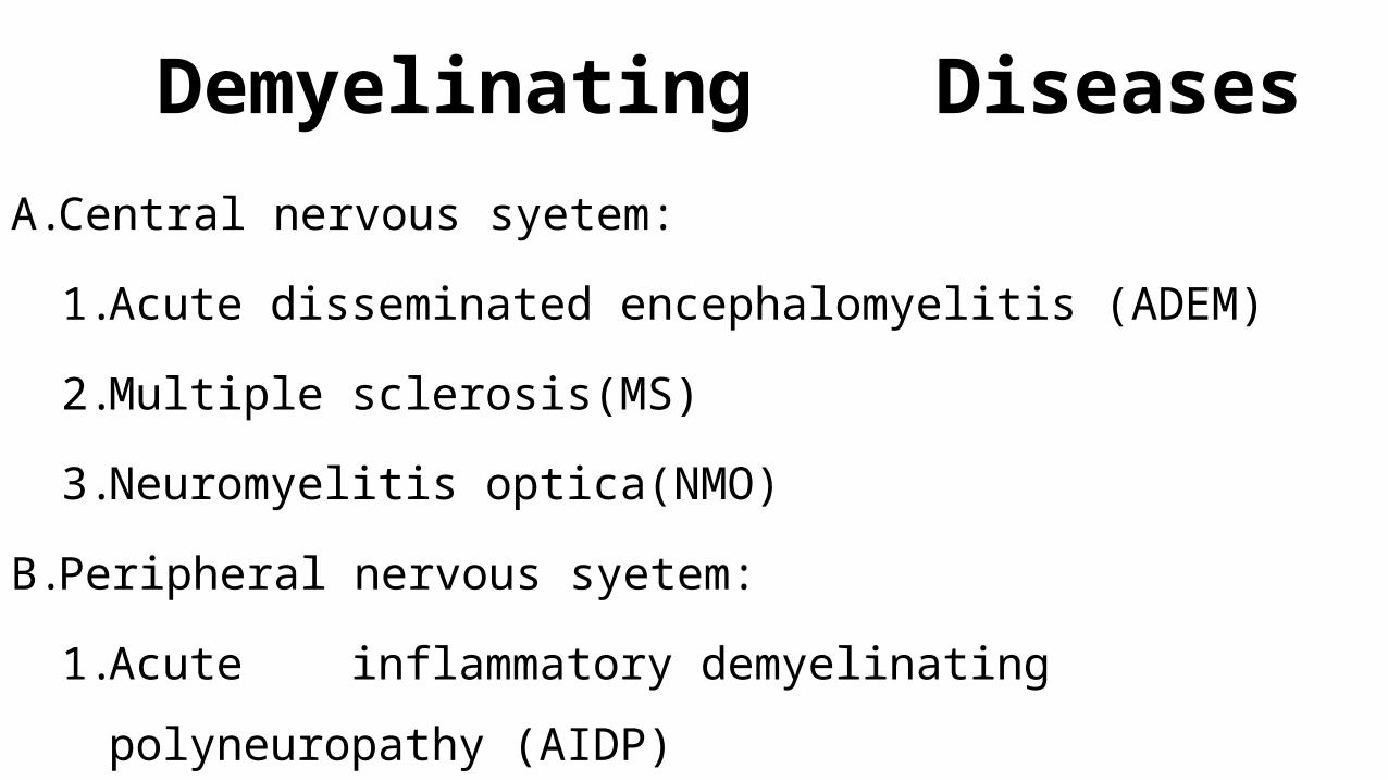
Demyelinating DiseasesA. Central nervous syetem:
1. Acute disseminated encephalomyelitis (ADEM)
2. Multiple sclerosis(MS)
3. Neuromyelitis optica(NMO)
B. Peripheral nervous syetem:
1. Acute inflammatory demyelinating polyneuropathy (AIDP)
Definitionis a demyelinating disease of the central nervous system
that typically presents as
1. monophasic disorder
2. encephalopathy
3. multifocal neurologic symptoms
INTRODUCTION
• Acute Disseminated Encephalomyelitis (ADEM)
– Usually monophasic
– Demyelinating disorder
– Characterized by diffuse neurologic signs and symptoms
(polysymptomatic)
– Nuroimaging ; multifocal lesions of demyelination
EPIDEMIOLOGY ADEM is an uncommon illness
Mean age between 5 and 8 yr
estimated incidence is 0.4/100,000 population per year
Approximately three to six cases are seen each year
There is no specific ethnic distribution
indicate a slight male predominance
A seasonal distribution has been observed showing that most ADEM
cases occur in the winter and spring
PATHOGENESIS Most of these conditions are thought to be
– caused by immune system dysregulation
– triggered by an infectious or other environmental agent
– genetically susceptible host.

PATHOGENESISPostinfectious
Postvaccinial
Thought to occur from a cross reactivity in immunity
to viral antigens.
PATHOGENESISThe immunopathological events leading to ADEM
can be divided into two major phases :-
1. Initial T cell priming and activation
2. Subsequent recruitment and effector phase
PATHOGENESIS typically following a recent (1-2 weeks prior) viral infection
or vaccination
• Sites of demyelination ;
1. white matter
2. Gray matter, especially that of the basal ganglia, is also often
involved
3. lesser extent, as is the spinal cord.
PATHOGENESIS• Large number of viruses associated with these infections ;
– approximately 50-75 percent of ADEM cases
– including :
• Measles , Mumps, Rubella
• Varicella zoster, Epstein-Barr, Cytomegalovirus
• Herpes simplex, Hepatitis A, Influenza
• Enterovirus infections.
PATHOGENESIS• Less than 5 percent of ADEM cases follow immunization.
• Associated with immunization for:
• Rabies, Hepatitis B, Influenza
• Japanese B encephalitis
• Diphtheria /Pertussis / Tetanus
• Measles, Mumps , Rubella,
• Pneumococcus, Polio, Smallpox, and Varicella.
PATHOGENESIS
• measles, mumps, and rubella vaccinations are most
commonly associated with post- vaccinial ADEM.

CLINICAL FEATURES
• Initial Presentation:
1. Fever,
2. Headache,
3. Vomiting
4. meningismus
CLINICAL FEATURES • febrile illness occurs in 50 to 75 percent of children
• Neurologic symptoms typically appear 4 to 13 days after the
infection or vaccination
CLINICAL FEATURES
• The Neurological Signs :
1. Encephalopathy ,
2. multifocal neurologic deficits
CLINICAL FEATURES • Encephalopathy
• is a characteristic feature of ADEM
• usually develops rapidly.
CLINICAL FEATURES • Encephalopathy results in symptoms, such as:
1. Altered level of consciousness
I. lethargy →coma
II. Confusion
III. Excessive irritability
2. Acute cognitive dysfunction
3. Behavioral changes
4. Seizures
• In about ⅓ of those diagnosed
CLINICAL FEATURES • Other common neurologic signs of ADEM include:
1. Long tract pyramidal signs
2. Acute hemiparesis
3. Cerebellar ataxia
4. Cranial neuropathies
5. cranial neuropathies including optic neuritis
6. spinal cord dysfunction (transverse myelitis)
CLINICAL FEATURES • less commonneurologic signs of ADEM include:
1. Aphasia
2. movement disorders
3. sensory deficits
CLINICAL FEATURES • New clinical symptoms may develop during
hospitalization.
• Progression of neurologic signs to maximum deficits
usually occurs over four to seven days
CLINICAL FEATURES ; optic neuritis• Symptoms of optic neuritis include
1. vision loss
2. pain with eye movement
3. afferent pupillary defect.
CLINICAL FEATURES Transverse Myelitis
• Symptoms of transverse myelitis include
1. flaccid paralysis of the legs
2. sensory level on examination.
• The arms can be involved if the demyelinating lesion is in the cervical cord.
• Respiratory failure may occur with high cervical lesions that extend into the brainstem.
– Bowel and bladder involvement secondary to spinal cord disease results in
constipation and urinary retention
Clinical course
• The severe phase of ADEM typically lasts from two to four weeks.
• Children may deteriorate after hospital admission
– many develop new neurologic signs.
• Patients usually recover completely from the acute illness
– although some have neurologic sequelae.
hyperacute variants of ADEM Inflammatory hemorrhagic demyelination of central nervous system white
matter is seen in rare conditions that are considered to be hyperacute
variants of ADEM
hyperacute variants of ADEM o These include:
1.Acute hemorrhagic leukoencephalitis (AHL)
2.Acute hemorrhagic encephalomyelitis (AHEM)
3.Acute necrotizing hemorrhagic leukoencephalitis (ANHLE) of
Weston Hurst
Acute hemorrhagic leukoencephalitis • These hemorrhagic variants are more rapidly progressive and more severe
than typical ADEM.
• symptomatology is similar to ADEM, with meningismus, headache, seizures,
multifocal neurologic signs, asymmetrical neurologic deficits, and coma.
• They typically follow an upper respiratory infection. (similar to ADEM)
Brain imaging with MRI Acute hemorrhagic leukoencephalitis
o diffuse white matter lesions, often large
o associated with cerebral edema
Brain imaging with MRI Acute hemorrhagic leukoencephalitis
o hemorrhage itself is not necessarily seen with conventional
T2-weighted and fluid-attenuated inversion recovery
(FLAIR) sequences.
o spin-echo MRI sequences are more readily able to identify
the acute hemorrhage associated with this form of ADEM.
Acute hemorrhagic leukoencephalitis
• Some patients recover with treatment.
• However, the prognosis for survival and recovery of
neurologic function is worse for AHL than ADEM.

Laboratory studies
• nonspecific findings of inflammation.
– Leukocytosis is common, occurring in up to two-thirds of patients
• predominantly ; lymphocytosis
– Platelet counts are elevated in a number of children with ADEM.
– C-reactive protein concentration may be increased.
– Sedimentation rates are elevated in a third of patients

Investigation• Investigation for infectious agents usually includes
– viral cultures of the throat and nasopharynx, stool, and CSF
– serologic testing for a variety of agents, including influenza, Epstein-Barr
virus, herpes, varicella, mycoplasma, cytomegalovirus, and rubella.
– These studies are rarely positive
The cerebrospinal fluid• The cerebrospinal fluid typically shows both ;
– Cerebrospinal fluid (CSF) is abnormal in about two-thirds of patients
– CSF can also be normal
• Evidence of inflammation
1. white and red blood cells
2. increased protein concentration
3. increase in myelin basic protein.
• CSF myelin basic protein concentration level, reflecting demyelination, is frequently elevated
in ADEM.
Lumbar puncture • Some patients with ADEM have oligoclonal bands in CSF.
• Oligoclonal bands
– are a nonspecific finding more often associated with multiple sclerosis;
– they may also occur in
1. chronic central nervous system infections
2. viral syndromes
3. neuropathies.
The cerebrospinal fluid
• Results of CSF immune profile testing (eg, CSF:serum
immunoglobulin G [IgG] index, CNS IgG synthetic rate,
oligoclonality) employing age-appropriate normative data
are positive in fewer than 10% of prepubertal children
with ADEM
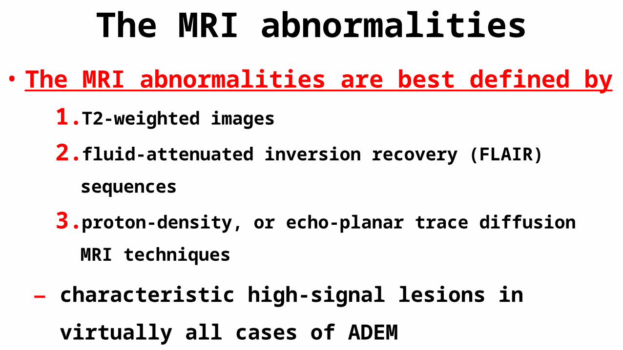
The MRI abnormalities• The MRI abnormalities are best defined by
1. T2-weighted images
2. fluid-attenuated inversion recovery (FLAIR) sequences
3. proton-density, or echo-planar trace diffusion MRI techniques
– characteristic high-signal lesions in virtually all cases of
ADEM
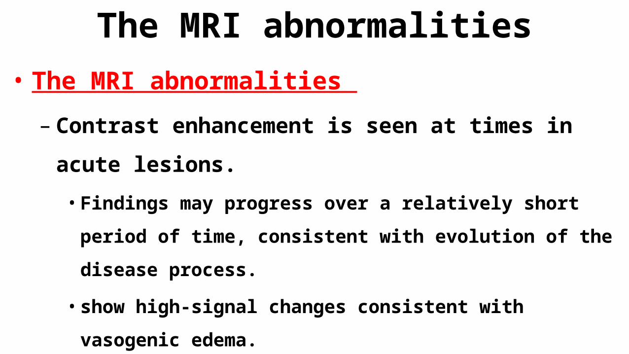
The MRI abnormalities• The MRI abnormalities
– Contrast enhancement is seen at times in acute lesions.
• Findings may progress over a relatively short period of time,
consistent with evolution of the disease process.
• show high-signal changes consistent with vasogenic edema.
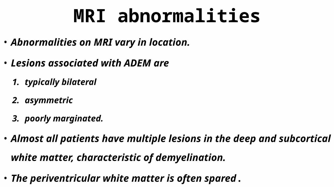
MRI abnormalities• Abnormalities on MRI vary in location.
• Lesions associated with ADEM are
1. typically bilateral
2. asymmetric
3. poorly marginated.
• Almost all patients have multiple lesions in the deep and subcortical white
matter, characteristic of demyelination.
• The periventricular white matter is often spared.
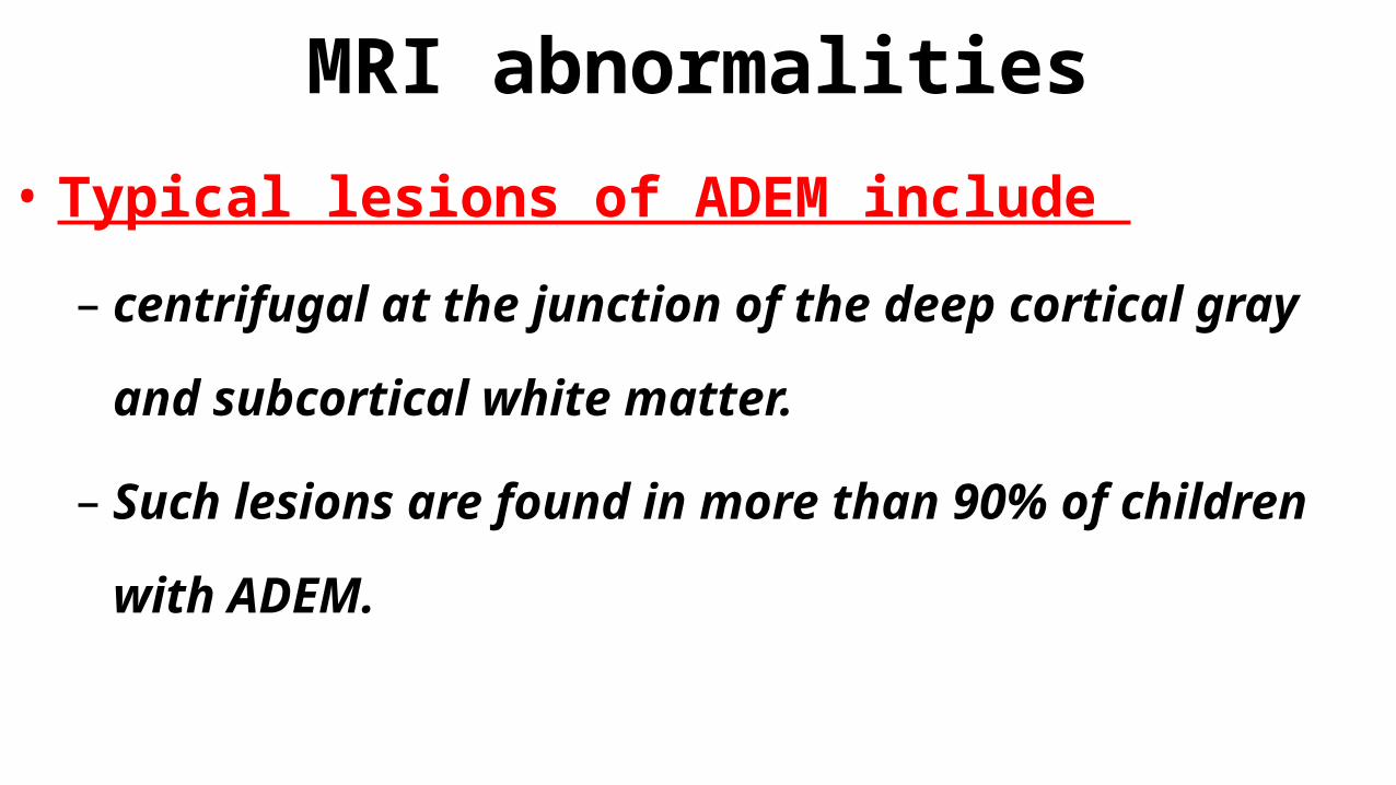
MRI abnormalities• Typical lesions of ADEM include
– centrifugal at the junction of the deep cortical gray and
subcortical white matter.
– Such lesions are found in more than 90% of children with
ADEM.
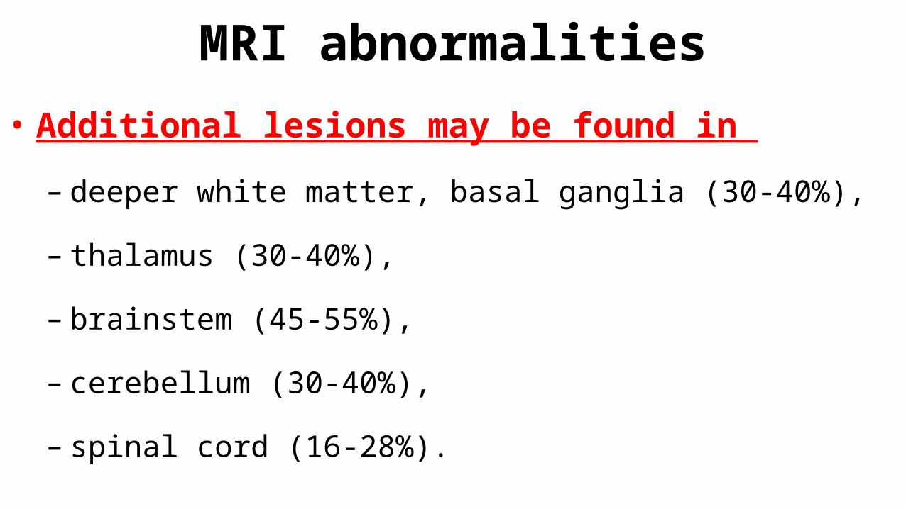
MRI abnormalities• Additional lesions may be found in
– deeper white matter, basal ganglia (30-40%),
– thalamus (30-40%),
– brainstem (45-55%),
– cerebellum (30-40%),
– spinal cord (16-28%).
MRI abnormalities• Periventricular lesions and corpus callosal lesions are
uncommon in childhood ADEM compared with MS
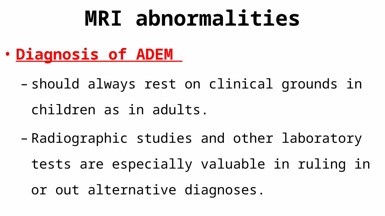
MRI abnormalities• Diagnosis of ADEM
– should always rest on clinical grounds in children as in adults.
– Radiographic studies and other laboratory tests are especially
valuable in ruling in or out alternative diagnoses.
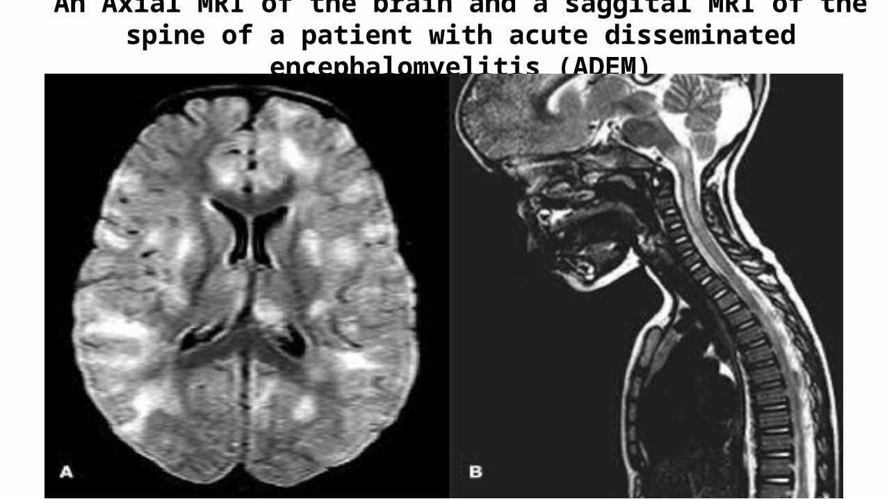
An Axial MRI of the brain and a saggital MRI of the spine of a patient with acute disseminated encephalomyelitis (ADEM)
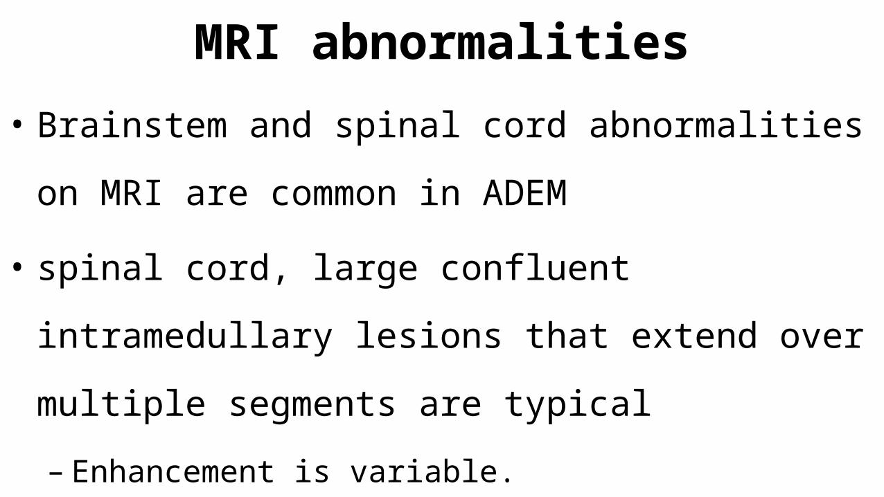
MRI abnormalities• Brainstem and spinal cord abnormalities on MRI are
common in ADEM
• spinal cord, large confluent intramedullary lesions that
extend over multiple segments are typical
– Enhancement is variable.
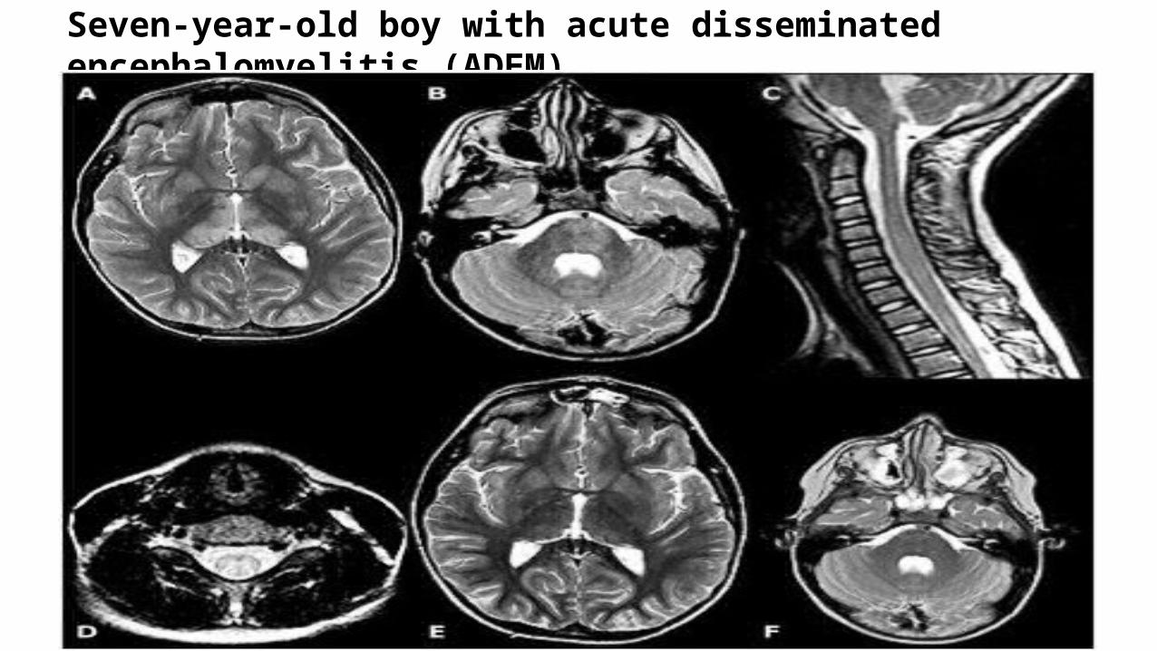
Seven-year-old boy with acute disseminated encephalomyelitis (ADEM)
MRI
• Magnetization transfer
– may help distinguish ADEM from MS, in that normal appearing brain (on T2
weighted images) has normal magnetization transfer ratio (MTR) and
normal diffusivity,
– whereas in MS both measurements are significantly decreased
CT
• The CT scan
– low-density abnormalities
– found in more than half of childhood or adolescent ADEM cases
– but this technique is far less sensitive than MRI for the
disclosure of extent and number of lesions.
electroencephalogram (EEG)
• The electroencephalogram (EEG) is not diagnostic
– It may show background slow wave activity that is typical of an
encephalopathy
– Seizure activity is seen.
visual-evoked potentials
• In patients with optic neuritis, visual-evoked potentials
may be prolonged.
DIAGNOSIS• The diagnosis of ADEM is based upon the
1. clinical
2. radiologic features
– There is no specific biologic marker or confirmatory test.
DIAGNOSIS• An ADEM diagnosis :is considered when – individuals develop multifocal neurologic abnormalities with:
1. Confusion
2. Excessive irritability
3. Altered level of consciousness (encephalopathy)
• Especially if the onset of symptoms occurs within 1 to 2 weeks after
a viral/bacterial infection or a vaccination
DIAGNOSIS
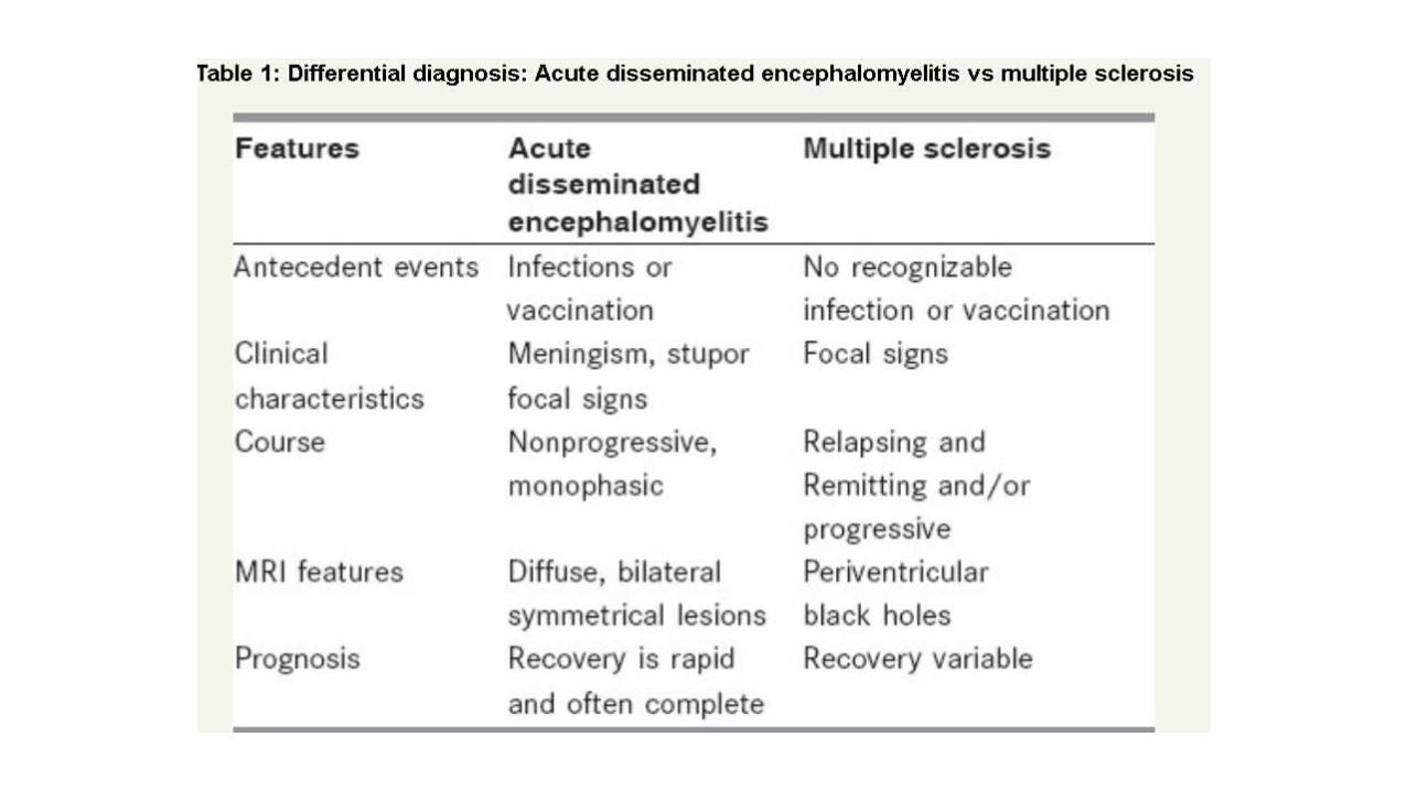
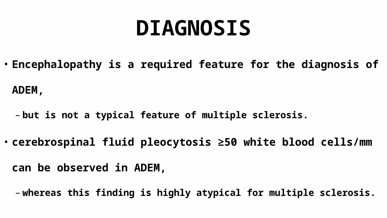
• Encephalopathy is a required feature for the diagnosis of ADEM,
– but is not a typical feature of multiple sclerosis.
• cerebrospinal fluid pleocytosis ≥50 white blood cells/mm can be
observed in ADEM,
– whereas this finding is highly atypical for multiple sclerosis.

DIAGNOSIS
• Bacterial and viral meningitis or encephalitis must be considered
and ruled out.
• Empiric treatment with broad-spectrum antibiotics and
acyclovir should be considered until an infectious etiology is
excluded
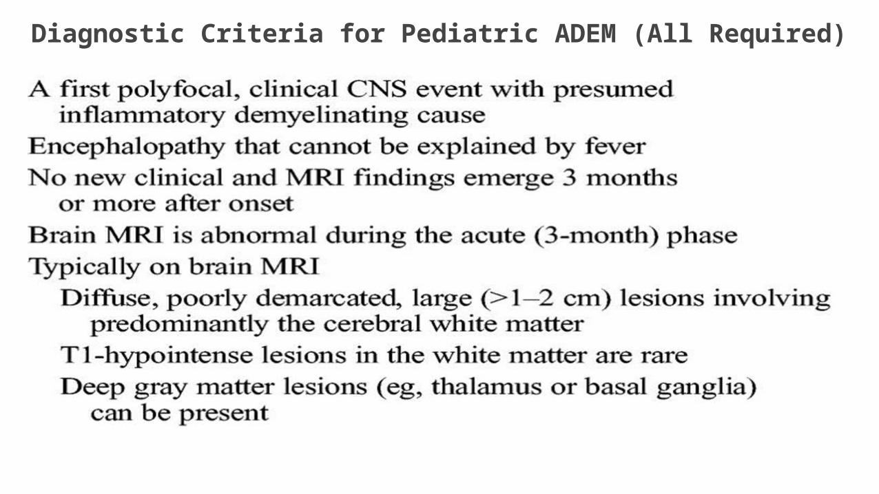
Diagnostic Criteria for Pediatric ADEM (All Required)
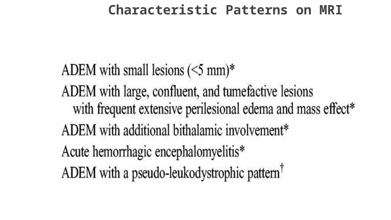
Characteristic Patterns on MRI
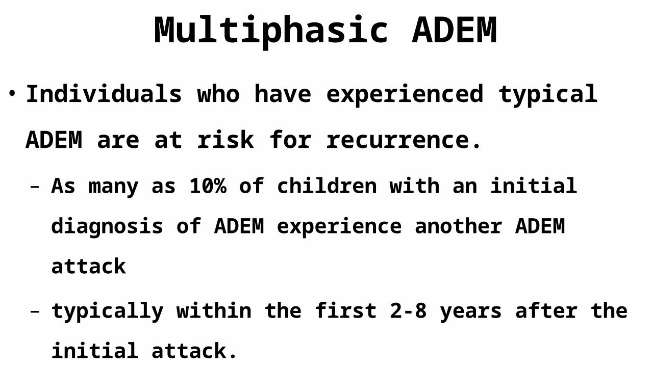
Multiphasic ADEM
• Individuals who have experienced typical ADEM are at
risk for recurrence.
– As many as 10% of children with an initial diagnosis of ADEM
experience another ADEM attack
– typically within the first 2-8 years after the initial attack.
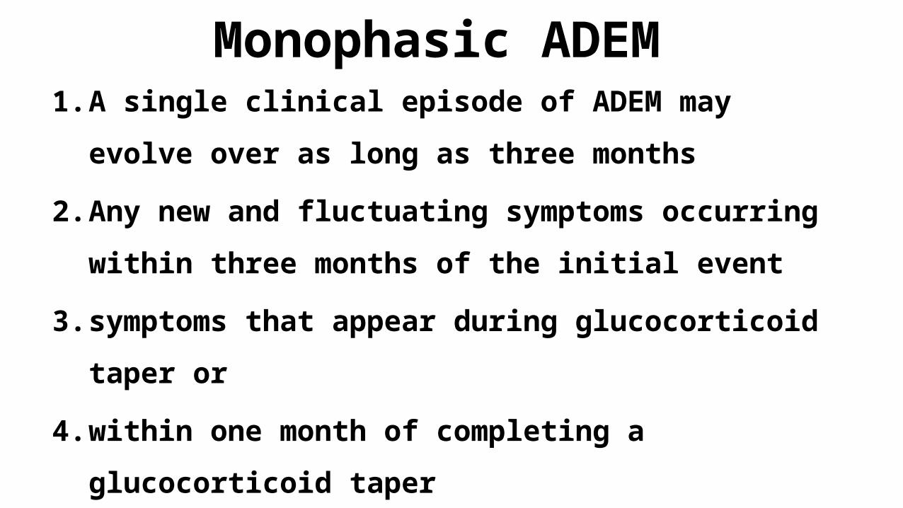
Monophasic ADEM 1. A single clinical episode of ADEM may evolve over as
long as three months
2. Any new and fluctuating symptoms occurring within
three months of the initial event
3. symptoms that appear during glucocorticoid taper or
4. within one month of completing a glucocorticoid taper
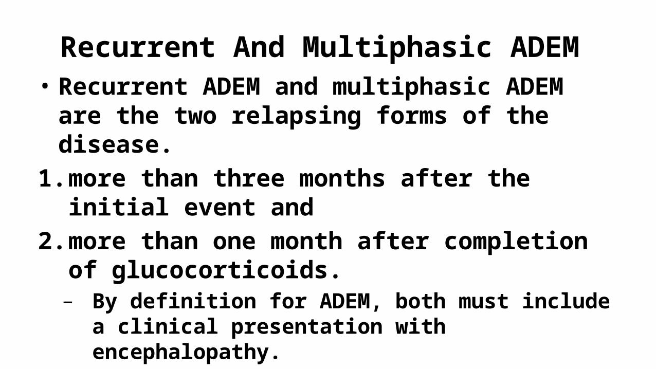
Recurrent And Multiphasic ADEM • Recurrent ADEM and multiphasic ADEM are the
two relapsing forms of the disease. 1. more than three months after the initial event
and 2. more than one month after completion of
glucocorticoids. – By definition for ADEM, both must include a clinical
presentation with encephalopathy.
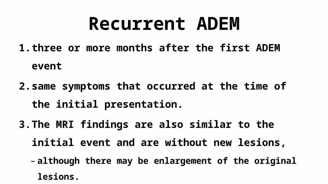
Recurrent ADEM1. three or more months after the first ADEM event
2. same symptoms that occurred at the time of the
initial presentation.
3. The MRI findings are also similar to the initial event
and are without new lesions,
– although there may be enlargement of the original lesions.
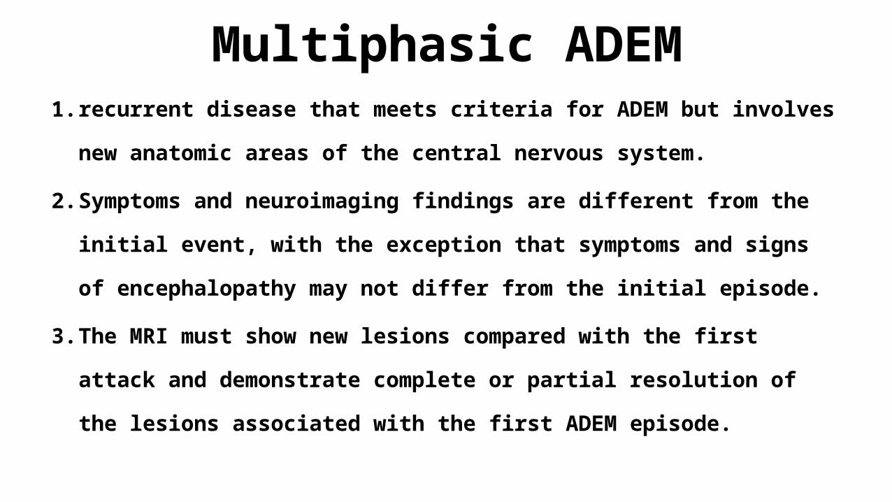
Multiphasic ADEM1. recurrent disease that meets criteria for ADEM but involves new
anatomic areas of the central nervous system.
2. Symptoms and neuroimaging findings are different from the initial event,
with the exception that symptoms and signs of encephalopathy may not
differ from the initial episode.
3. The MRI must show new lesions compared with the first attack and
demonstrate complete or partial resolution of the lesions associated
with the first ADEM episode.
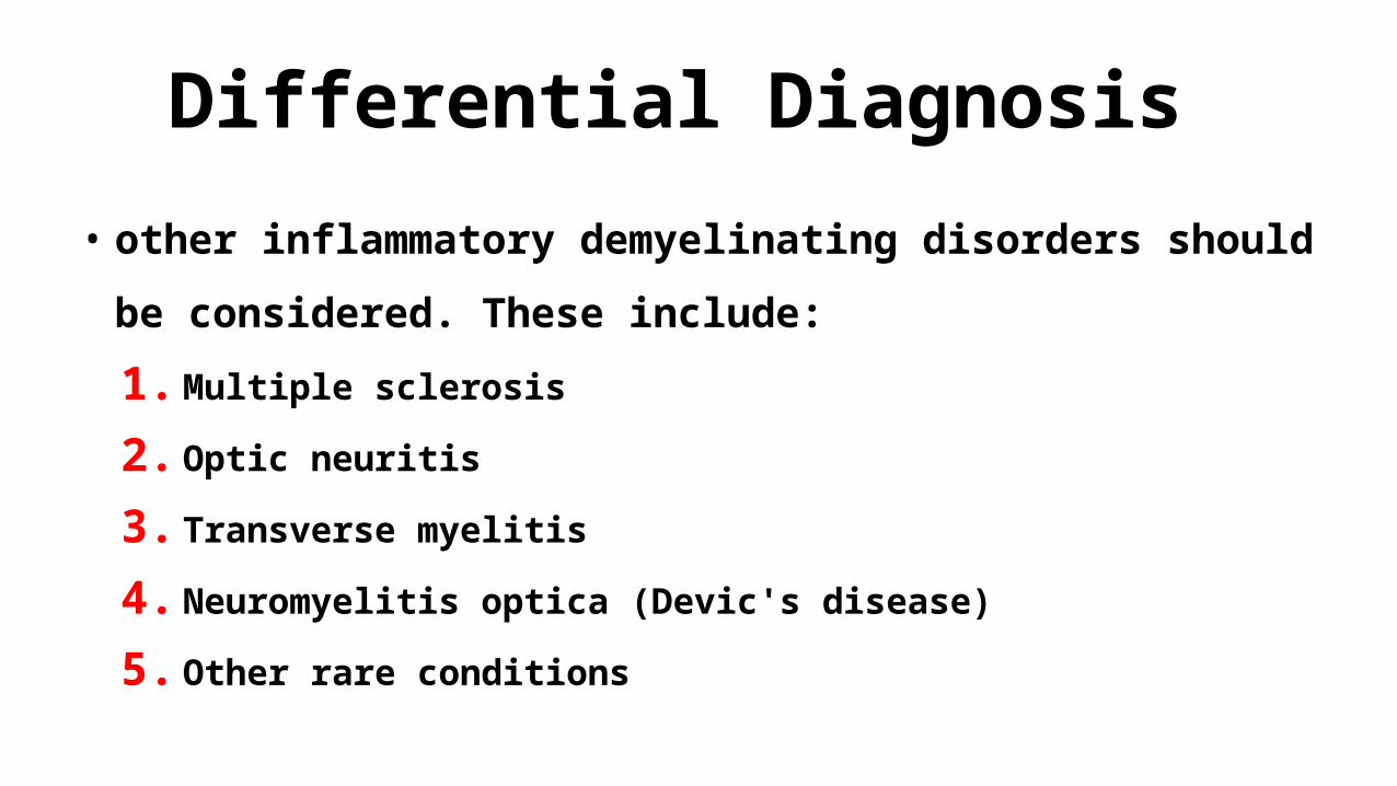
Differential Diagnosis • other inflammatory demyelinating disorders should be considered.
These include:
1. Multiple sclerosis
2. Optic neuritis
3. Transverse myelitis
4. Neuromyelitis optica (Devic's disease)
5. Other rare conditions
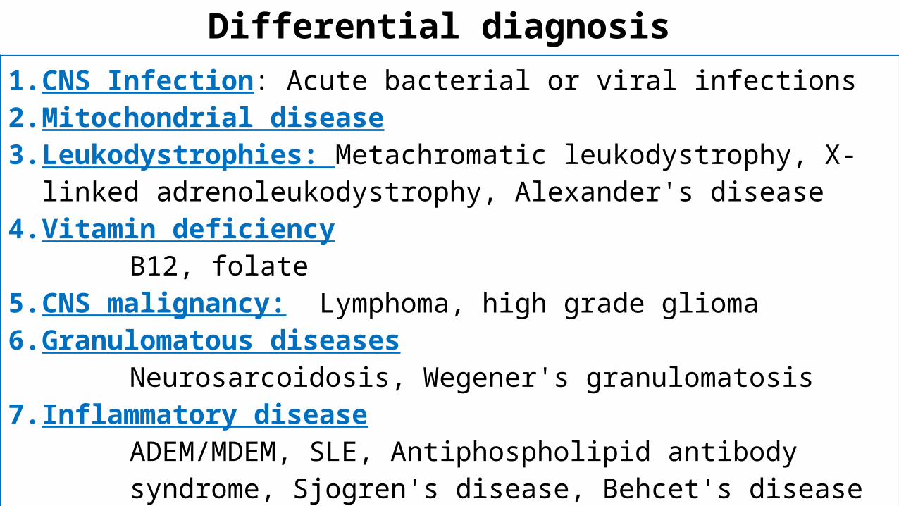
Differential diagnosis 1.CNS Infection: Acute bacterial or viral infections2.Mitochondrial disease3.Leukodystrophies: Metachromatic leukodystrophy, X-linked
adrenoleukodystrophy, Alexander's disease4.Vitamin deficiency
B12, folate 5.CNS malignancy: Lymphoma, high grade glioma6.Granulomatous diseases
Neurosarcoidosis, Wegener's granulomatosis7. Inflammatory disease
ADEM/MDEM, SLE, Antiphospholipid antibody syndrome, Sjogren's disease, Behcet's disease
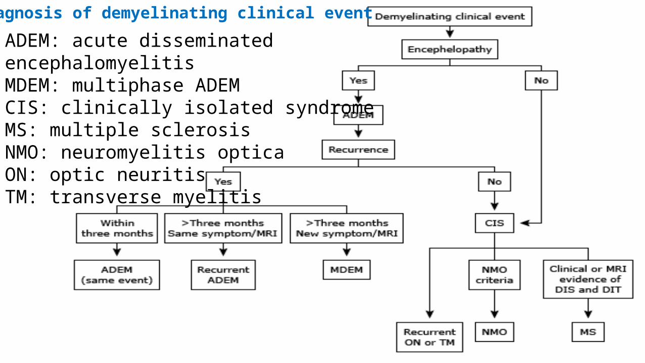
Diagnosis of demyelinating clinical event
ADEM: acute disseminated encephalomyelitisMDEM: multiphase ADEMCIS: clinically isolated syndromeMS: multiple sclerosisNMO: neuromyelitis opticaON: optic neuritisTM: transverse myelitis
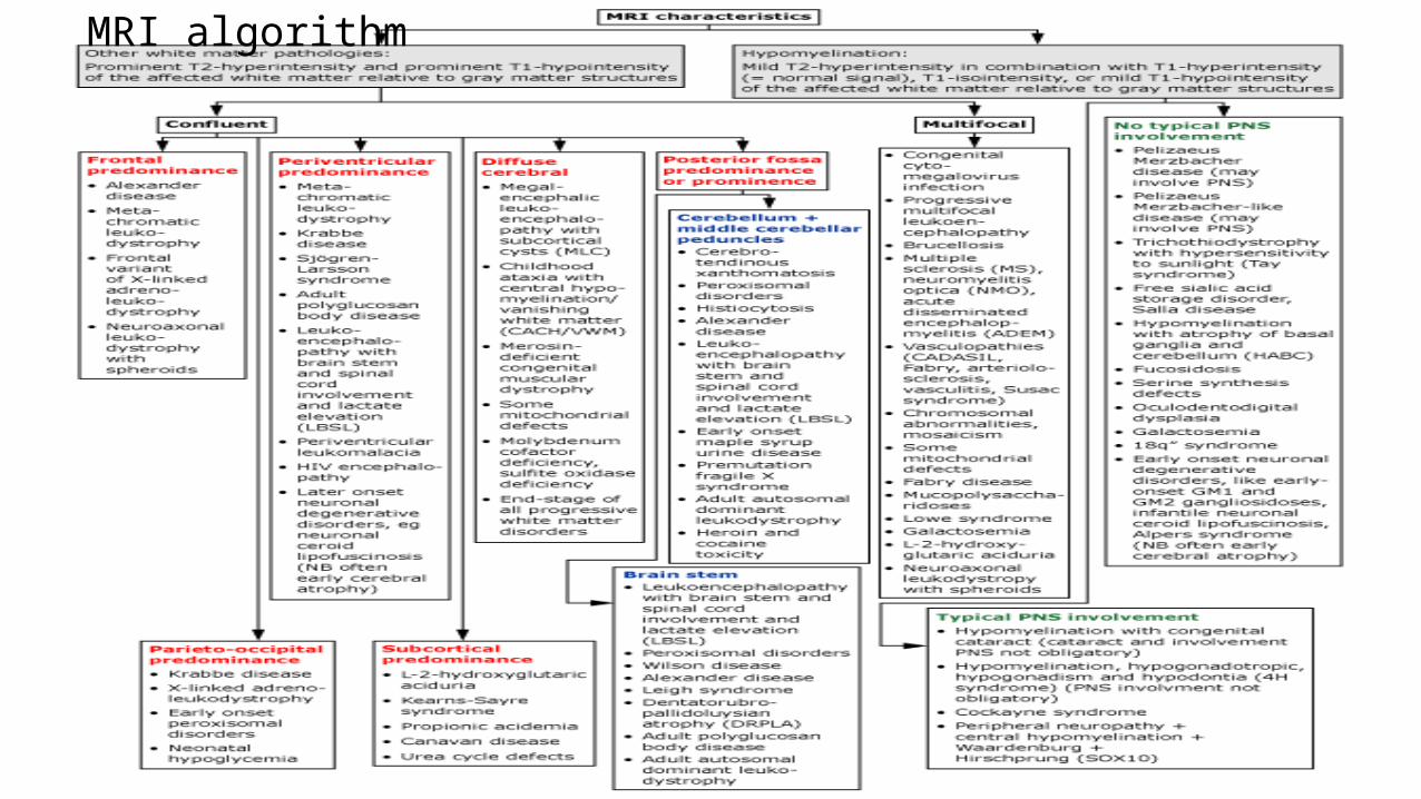
MRI algorithm
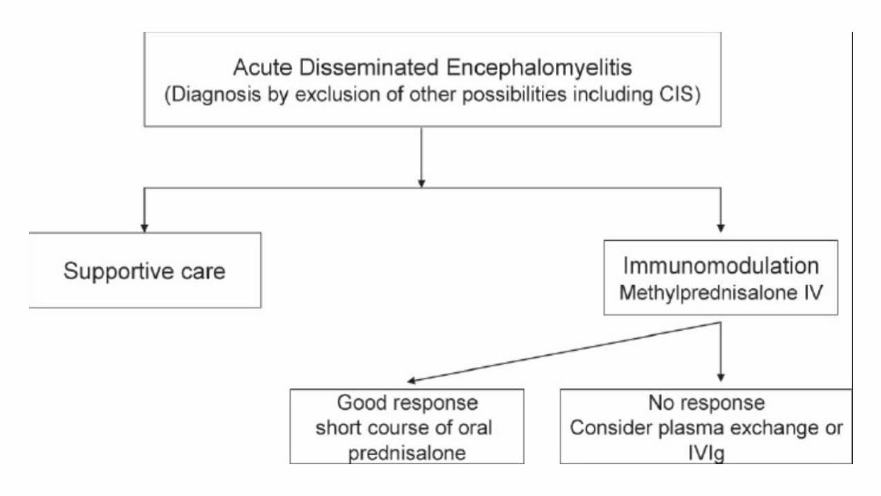
TREATMENT
• It is important to first consider a treatment with
antibiotics and/or acyclovir until an infectious cause is
ruled out
• A high dose of intravenous corticosteroids, for 3-5 days
is the primary and most common first treatment of
ADEM
TREATMENT1. high-dose intravenous (IV) glucocorticoids. 2. intravenous immune globulin 3. plasma exchange
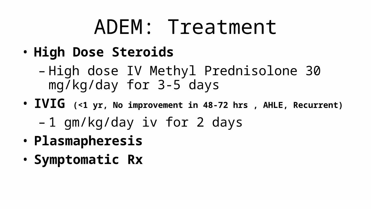
ADEM: Treatment• High Dose Steroids– High dose IV Methyl Prednisolone 30 mg/kg/day for 3-5
days • IVIG (<1 yr, No improvement in 48-72 hrs , AHLE, Recurrent)
– 1 gm/kg/day iv for 2 days• Plasmapheresis• Symptomatic Rx
TREATMENT• For children with ADEM, we recommend
immunosuppressive treatment
• We suggest initial therapy with high-dose glucocorticoids
• IV methylprednisolone (30 mg/kg per day, up to a
maximum dose of 1000 mg per day) for five days without
a taper.

TREATMENT
• For children with ADEM who have an insufficient response to IV
glucocorticoid treatment, we suggest
intravenous immune globulin treatment
• For children with ADEM who do not respond to glucocorticoids
and IVIG, we suggest treatment with plasma exchange
PROGNOSIS• Prognosis for most children with ADEM is good
• Recovery is usually a slow process lasting from four to
six weeks
• The majority of children with ADEM make a full
recovery
PROGNOSIS
• Between 60 to 90 percent are left with no neurological deficits
• Those children who do have residual symptoms are reported to have symptoms
from:
• Transverse myelitis
• Recurrent headaches
• Behavioral problems
PROGNOSIS
• Long-term clinical follow-up and sequential imaging by
MRI are normally required to confirm a diagnosis of
ADEM.
PROGNOSIS
• Development of a relapse with new lesions, it is not
compatible with a diagnosis of monophasic ADEM

PROGNOSIS
• Depending on the clinical and imaging features, it likely
suggests the correct diagnosis being either multiphasic
ADEM or MS.
PROGNOSIS
• Depending on the clinical and imaging features, it likely
suggests the correct diagnosis being either multiphasic
ADEM or MS.
PROGNOSIS
• The prognosis for survival and recovery of
neurologic function is worse for the hyperacute
hemorrhage variants of ADEM, such as acute
hemorrhagic leukoencephalitis, than for typical
ADEM
PROGNOSIS• Complete recovery in 10 (77%) of the survivors• Relapsing disease in 2 (15%)• Mortality less than 2%– fulminant cervical transverse myelitis or brain
swelling. Children younger than 2 years
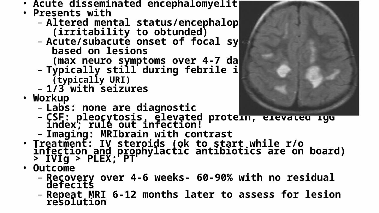
• Acute disseminated encephalomyelitis• Presents with
– Altered mental status/encephalopathy(irritability to obtunded)
– Acute/subacute onset of focal symptoms based on lesions (max neuro symptoms over 4-7 days)
– Typically still during febrile illness (typically URI)
– 1/3 with seizures• Workup
– Labs: none are diagnostic– CSF: pleocytosis, elevated protein, elevated IgG index; rule out
infection!– Imaging: MRIbrain with contrast
• Treatment: IV steroids (ok to start while r/o infection and prophylactic antibiotics are on board) > IVIg > PLEX; PT
• Outcome– Recovery over 4-6 weeks- 60-90% with no residual defecits– Repeat MRI 6-12 months later to assess for lesion resolution