humanmilkoligosaccharidesinhibitgrowthofgroupb...
TRANSCRIPT
Human milk oligosaccharides inhibit growth of group BStreptococcusReceived for publication, April 6, 2017 Published, Papers in Press, April 17, 2017, DOI 10.1074/jbc.M117.789974
Ann E. Lin‡, Chloe A. Autran§¶, Alexandra Szyszka§¶, Tamara Escajadillo‡, Mia Huang!, Kamil Godula!,Anthony R. Prudden**, Geert-Jan Boons**, Amanda L. Lewis‡‡, Kelly S. Doran‡§§, Victor Nizet‡¶¶!!,and Lars Bode§¶!!***1
From the Divisions of ‡Host-Microbe Systems and Therapeutics, §Neonatology, and ¶Gastroenterology, Hepatology, and Nutrition,Department of Pediatrics, !Department of Chemistry and Biochemistry, ¶¶Skaggs School of Pharmacy and PharmaceuticalSciences, and ***Larsson-Rosenquist Foundation Mother-Milk-Infant Center of Research Excellence, University of California, SanDiego, La Jolla, California 92093, !!Rady Children’s Hospital, San Diego, California 92123, the **Complex Carbohydrate ResearchCenter, University of Georgia, Athens, Georgia 30602, the ‡‡Department of Molecular Microbiology and Center for Women’sInfectious Disease Research, Washington University School of Medicine, St. Louis, Missouri 63110, and the §§Department of Biologyand Center for Microbial Sciences, San Diego State University, San Diego, California 92182
Edited by Gerald W. Hart
Streptococcus agalactiae (group B Streptococcus, GBS) is aleading cause of invasive bacterial infections in newborns, typi-cally acquired vertically during childbirth secondary to mater-nal vaginal colonization. Human milk oligosaccharides (HMOs)have important nutritional and biological activities that guidethe development of the immune system of the infant and shapethe composition of normal gut microbiota. In this manner,HMOs help protect against pathogen colonization and reducethe risk of infection. In the course of our studies of HMO-micro-bial interactions, we unexpectedly uncovered a novel HMOproperty to directly inhibit the growth of GBS independent ofhost immunity. By separating different HMO fractions throughmultidimensional chromatography, we found the bacteriostaticactivity to be confined to specific non-sialylated HMOs and syn-ergistic with a number of conventional antibiotic agents. Pheno-typic screening of a GBS transposon insertion library identifieda mutation within a GBS-specific gene encoding a putative gly-cosyltransferase that confers resistance to HMOs, suggestingthat HMOs may function as an alternative substrate to modify aGBS component in a manner that impairs growth kinetics. Ourstudy uncovers a unique antibacterial role for HMOs against aleading neonatal pathogen and expands the potential therapeu-tic utility of these versatile molecules.
Group B Streptococcus (GBS)2 are Gram-positive bacteriathat colonize the vaginal epithelium in 15–30% of healthy
women. GBS transmission to the newborn is associated withrisk of pneumonia, septicemia, and meningitis (1–3). In theUnited States and other developed countries, implementationof universal antenatal GBS culture screening and administra-tion of intrapartum antibiotic prophylaxis has reduced GBSincidence in the first few days of life; however, it has not had asimilar impact on late-onset infections, which now representapproximately one-third of total cases (4). Up to half of allinfants with late-onset GBS also develop meningitis, which car-ries a high incidence (!40%) of neurocognitive sequelae amongsurvivors (5). In a recent meta-analysis, the overall incidence ofGBS infection in infants !3 months of age in the Americas andEurope is "0.53– 0.67 cases/1000 births, with an overall casefatality rate of 7–10% (6). The emergence of antibiotic-resistantGBS strains has become an increasing concern (1, 7, 8).
Human milk oligosaccharides (HMOs) are a group of com-plex carbohydrates that are highly abundant in human milk(10 –15 g/liter) but not in infant formula (reviewed in Ref. 9).HMOs are comprised of five monosaccharides: D-glucose (Glc),D-galactose (Gal), GlcNAc, L-fucose, and sialic acid (N-acetyl-neuraminic acid). The Gal-Glc disaccharide (lactose) backbonecan be further elongated by up to 15 Gal-GlcNAc repeats andcan be sialylated or fucosylated. Over 150 structurally dis-tinct HMOs have been identified, comprised of neutral (non-sialylated) and acidic (sialylated) forms, and the amount andcomposition are highly variable between women (reviewedin Ref. 10).
HMOs are not digested by the infant and reach the colonintact, where they serve as metabolic substrates for specific,potentially beneficial bacteria and help shape the infant micro-biome. HMOs also act as soluble receptor decoys to preventattachment of microbial pathogens to the host (11–14). HMOsare partially absorbed and reach the systemic circulation of the
This work was supported by NHLBI, National Institutes of Health Programs ofExcellence in Glycosciences Grant HL107150 (to V. N.), NIDDK, NationalInstitutes of Health Grant P50DK064540 (PI: Hultgren) (to A. L. L.), and aPROMO fellowship from the German Academic Exchange Service (to A. S.).The authors declare that they have no conflicts of interest with the con-tents of this article. The content is solely the responsibility of the authorsand does not necessarily represent the official views of the National Insti-tutes of Health.
This article contains supplemental Figs. S1–S3.1 To whom correspondence should be addressed. Tel.: 858-246-1874; E-mail:
[email protected] The abbreviations used are: GBS, group B Streptococcus; HMO, human milk
oligosaccharide; Glc, D-glucose; Gal, D-galactose; UPEC, uropathogenicEscherichia coli; pHMO, pooled human milk oligosaccharide; GOS, galacto-oligosaccharide(s); aHMO, acidic human milk oligosaccharide; nHMO, neu-
tral human milk oligosaccharide; LNT, lacto-N-tetraose; LNnT, lacto-N-neotetraose; LNnH, lacto-N-neohexaose; LNFPI, lacto-N-fucopentaose I;LNDFHII, lacto-N-difucohexaose II; LnNO, lacto-N-neooctaose; LNnDFH,lacto-N-neodifucohexaose; LNnFPV, lacto-N-neofucopentaose; LNFPV,lacto-N-fucopentaose V; THB, Todd-Hewitt broth; SF-RPMI 1640, serum-free RPMI 1640; ANOVA, analysis of variance.
crosARTICLE
J. Biol. Chem. (2017) 292(27) 11243–11249 11243© 2017 by The American Society for Biochemistry and Molecular Biology, Inc. Published in the U.S.A.
at Biomedical Library, U
CSD on July 7, 2017
http://ww
w.jbc.org/
Dow
nloaded from
at Biomedical Library, U
CSD on July 7, 2017
http://ww
w.jbc.org/
Dow
nloaded from
at Biomedical Library, U
CSD on July 7, 2017
http://ww
w.jbc.org/
Dow
nloaded from
at Biomedical Library, U
CSD on July 7, 2017
http://ww
w.jbc.org/
Dow
nloaded from
at Biomedical Library, U
CSD on July 7, 2017
http://ww
w.jbc.org/
Dow
nloaded from
at Biomedical Library, U
CSD on July 7, 2017
http://ww
w.jbc.org/
Dow
nloaded from
infant (15, 16) and appear intact in the urine of breast-fedinfants (17, 18), with the potential to exert effects in organsother than the gut and including the urinary tract. Most intrigu-ingly, HMOs also appear in the urine of pregnant women asearly as the end of the first trimester (19), suggesting thatHMOs might already affect pregnant women and the growingfetus long before birth.
In a previous study, we demonstrated that HMOs regulatethe host innate immune response in bladder epithelial cellsto prevent invasion and cytotoxicity caused by uropathogenicEscherichia coli (UPEC) without any direct interference withbacterial growth (20). To delineate whether HMOs generatesimilar effects with other neonatal pathogens, we examinedthe effect of HMOs on GBS, which is commonly found in theurogenital tract of pregnant women. Unexpectedly, we foundthat HMOs directly inhibit the growth of GBS, a property notshared with UPEC, Pseudomonas aeruginosa, or Staphylo-coccus aureus. Further investigation revealed a unique aspectof HMOs that causes a significant GBS growth defect.
Results
Human milk oligosaccharides inhibit growth of group BStreptococcus
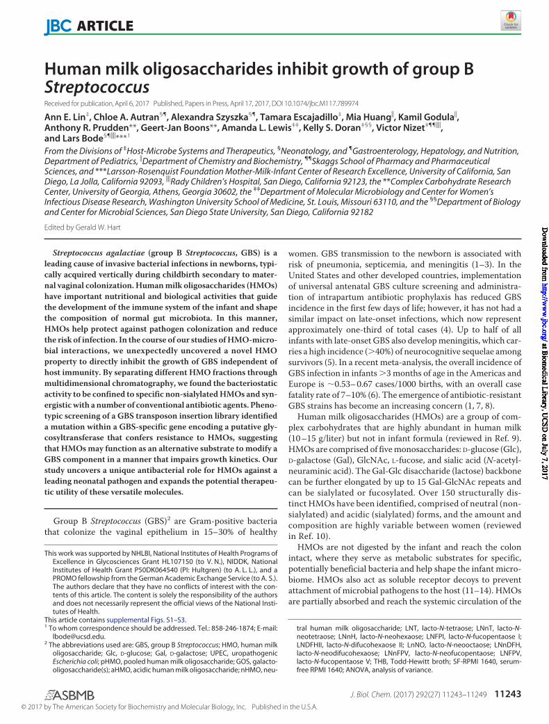
To evaluate the potential antimicrobial effect(s) of HMO ona group of bacterial pathogens, we resuspended "105 cfu ofeach bacterial overnight culture in serum-free tissue culturemedium (RPMI) with or without 2 mg/ml of HMOs isolatedfrom pooled human milk (pHMO) and then incubated for 4 h.pHMOs did not affect the growth of UPEC, P. aeruginosa, andmethicillin-resistant S. aureus. However, growth of the GBStest strain (serotype III isolate COH1) was reduced by "10-fold
(p # 0.05) (Fig. 1A). We confirmed that HMOs are bacterio-static and not bactericidal because they did not kill GBS even atvery high concentration (Fig. 1B). pHMO impaired the growthof the three most common GBS serotypes—serotypes III (strainCOH1), Ia (strain A909), and V (strain NCTC10/84)—in adose-dependent manner between 0.25–1.0 mg/ml (Fig. 1C).
The neutral fraction of HMOs possesses the GBS inhibitoryactivity
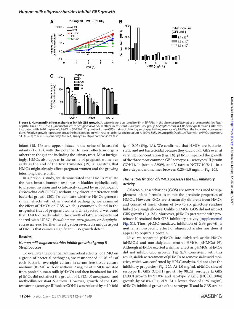
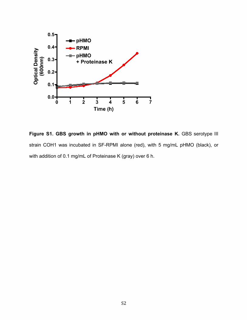
Galacto-oligosaccharides (GOS) are sometimes used to sup-plement infant formula to mimic the prebiotic properties ofHMOs. However, GOS are structurally different from HMOsand consist of linear chains of two to six galactose residueslinked to a single glucose. Unlike pHMOs, GOS did not impactGBS growth (Fig. 2A). Moreover, pHMOs pretreated with pro-teinase K retained their GBS-inhibitory activity (supplementalFig. S1). Thus, pHMO-mediated inhibition of GBS growth isneither a nonspecific effect of oligosaccharides nor does itappear to require a protein.
Next, we separated pHMOs into sialylated, acidic HMOs(aHMOs) and non-sialylated, neutral HMOs (nHMOs) (9).Although nHMOs exerted a similar effect as pHMOs, aHMOsdid not inhibit GBS growth (Fig. 2B). Consistent with thisresult, sialidase treatment of pHMOs to remove sialic acid moi-eties, which was confirmed by HPLC analysis, did not alter theinhibitory properties (Fig. 2C). At 1.0 mg/ml, nHMOs slowedserotype III GBS (COH1) growth by 98.2%, serotype Ia GBS(A909) growth by 97.0%, and serotype V GBS (NCTC10/84)growth by 96.0% (Fig. 2D). At a lower dose of 0.25 mg/ml,nHMOs inhibited growth of the serotype III and Ia GBS strains
Figure 1. Human milk oligosaccharides inhibit GBS growth. A, bacteria were cultured for 4 h in SF-RPMI in the absence (solid lines) or presence (dashed lines)of pHMO in a 37 °C, 5% CO2 incubator. Pa, P. aeruginosa; MRSA, methicillin-resistant S. aureus; GAS, group A Streptococcus. B, GBS serotype III strain COH1 wasincubated with 1–10 mg/ml of pHMO in SF-RPMI. C, growth of three GBS strains of differing serotypes in the presence of pHMOs at the indicated concentra-tions. Relative growth represents cfu at the indicated point with respect to initial cfu inoculum $ 100%. Solid line, no pHMOs; dotted line, with pHMOs; error bars,S.E. (n % 3). *, p # 0.05, one-way ANOVA, Tukey’s multiple comparison’s test.
Human milk oligosaccharides inhibit GBS growth
11244 J. Biol. Chem. (2017) 292(27) 11243–11249
at Biomedical Library, U
CSD on July 7, 2017
http://ww
w.jbc.org/
Dow
nloaded from
by more than 40-fold (p # 0.01), an effect more potent thanpHMOs at the same dose (Fig. 1C).
Identification of neutral HMOs that inhibit GBS growth
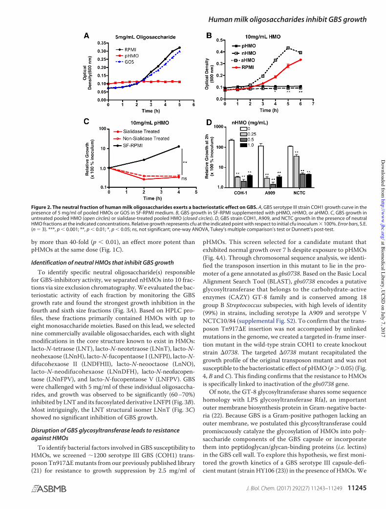
To identify specific neutral oligosaccharide(s) responsiblefor GBS-inhibitory activity, we separated nHMOs into 10 frac-tions via size exclusion chromatography. We evaluated the bac-teriostatic activity of each fraction by monitoring the GBSgrowth rate and found the strongest growth inhibition in thefourth and sixth size fractions (Fig. 3A). Based on HPLC pro-files, these fractions primarily contained HMOs with up toeight monosaccharide moieties. Based on this lead, we selectednine commercially available oligosaccharides, each with slightmodifications in the core structure known to exist in HMOs:lacto-N-tetraose (LNT), lacto-N-neotetraose (LNnT), lacto-N-neohexaose (LNnH), lacto-N-fucopentaose I (LNFPI), lacto-N-difucohexaose II (LNDFHII), lacto-N-neooctaose (LnNO),lacto-N-neodifucohexaose (LNnDFH), lacto-N-neofucopen-taose (LNnFPV), and lacto-N-fucopentaose V (LNFPV). GBSwere challenged with 5 mg/ml of these individual oligosaccha-rides, and growth was observed to be significantly (60 –70%)inhibited by LNT and its fucosylated derivative LNFPI (Fig. 3B).Most intriguingly, the LNT structural isomer LNnT (Fig. 3C)showed no significant inhibition of GBS growth.
Disruption of GBS glycosyltransferase leads to resistanceagainst HMOs
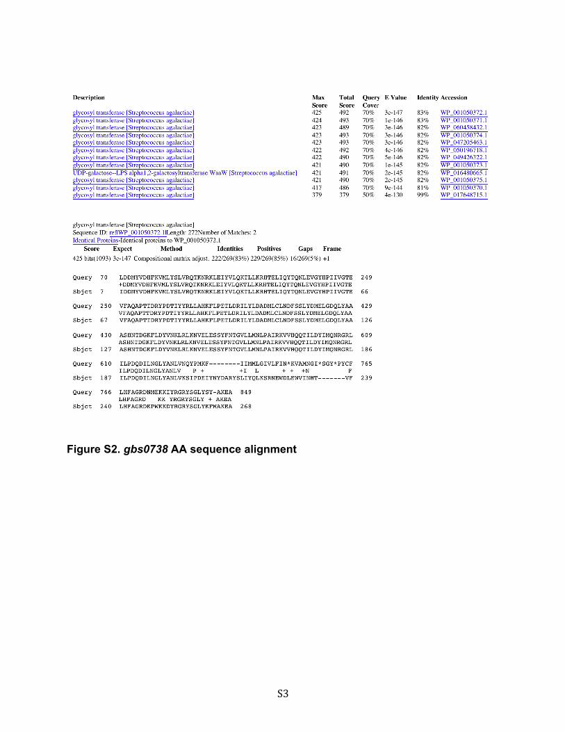
To identify bacterial factors involved in GBS susceptibility toHMOs, we screened "1200 serotype III GBS (COH1) trans-poson Tn917&E mutants from our previously published library(21) for resistance to growth suppression by 2.5 mg/ml of
pHMOs. This screen selected for a candidate mutant thatexhibited normal growth over 7 h despite exposure to pHMOs(Fig. 4A). Through chromosomal sequence analysis, we identi-fied the transposon insertion in this mutant to lie in the pro-moter of a gene annotated as gbs0738. Based on the Basic LocalAlignment Search Tool (BLAST), gbs0738 encodes a putativeglycosyltransferase that belongs to the carbohydrate-activeenzymes (CAZY) GT-8 family and is conserved among 18group B Streptococcus subspecies, with high levels of identity(99%) in strains, including serotype Ia A909 and serotype VNCTC10/84 (supplemental Fig. S2). To confirm that the trans-poson Tn917&E insertion was not accompanied by unlinkedmutations in the genome, we created a targeted in-frame inser-tion mutant in the wild-type strain COH1 to create knockoutstrain &0738. The targeted &0738 mutant recapitulated thegrowth profile of the original transposon mutant and was notsusceptible to the bacteriostatic effect of pHMO (p ! 0.05) (Fig.4, B and C). This finding confirms that the resistance to HMOsis specifically linked to inactivation of the gbs0738 gene.
Of note, the GT-8 glycosyltransferase shares some sequencehomology with LPS glycosyltransferase RfaJ, an importantouter membrane biosynthesis protein in Gram-negative bacte-ria (22). Because GBS is a Gram-positive pathogen lacking anouter membrane, we postulated this glycosyltransferase couldpromiscuously catalyze the glycosylation of HMOs into poly-saccharide components of the GBS capsule or incorporatethem into peptidoglycan/glycan-binding proteins (i.e. lectins)in the GBS cell wall. To explore this hypothesis, we first moni-tored the growth kinetics of a GBS serotype III capsule-defi-cient mutant (strain HY106 (23)) in the presence of HMOs. We
Figure 2. The neutral fraction of human milk oligosaccharides exerts a bacteriostatic effect on GBS. A, GBS serotype III strain COH1 growth curve in thepresence of 5 mg/ml of pooled HMOs or GOS in SF-RPMI medium. B, GBS growth in SF-RPMI supplemented with pHMO, nHMO, or aHMO. C, GBS growth inuntreated pooled HMO (open circles) or sialidase-treated pooled HMO (closed circles). D, GBS strain COH1, A909, and NCTC growth in the presence of neutralHMO fractions at the indicated concentrations. Relative growth represents cfu at the indicated point with respect to initial cfu inoculum $ 100%. Error bars, S.E.(n % 3). ***, p # 0.001; **, p # 0.01; *, p # 0.05; ns, not significant; one-way ANOVA, Tukey’s multiple comparison’s test or Dunnett’s post-test.
Human milk oligosaccharides inhibit GBS growth
J. Biol. Chem. (2017) 292(27) 11243–11249 11245
at Biomedical Library, U
CSD on July 7, 2017
http://ww
w.jbc.org/
Dow
nloaded from
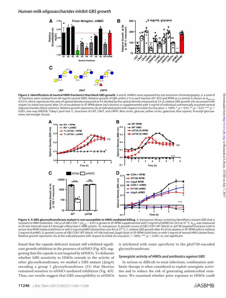
found that the capsule-deficient mutant still exhibited signifi-cant growth inhibition in the presence of nHMO (Fig. 4D), sug-gesting that the capsule is not targeted by nHMOs. To delineatewhether GBS sensitivity to HMOs extends to the activity ofother glycosyltransferases, we studied a GBS mutant (&iagA)encoding a group 1 glycosyltransferase (21) that likewiseremained sensitive to nHMO-mediated inhibition (Fig. 4D).Thus, our results suggest that GBS susceptibility to nHMOs
is attributed with some specificity to the gbs0738-encodedglycosyltransferase.
Synergistic activity of HMOs and antibiotics against GBS
In serious or difficult-to-treat infections, combination anti-biotic therapy is often considered to exploit synergistic activi-ties and to reduce the risk of generating antimicrobial resis-tance. We examined whether prior exposure to HMOs could
Figure 3. Identification of neutral HMO fraction(s) that block GBS growth. A and B, nHMOs were separated by size exclusion chromatography. A, a total of10 fractions were isolated from 40 mg/ml neutral HMO. Relative growth of GBS within 2 h in each fraction (N1-N10 and RPMI as a control) is shown as A600 nm4 h/2 h, which represents the ratio of optical density measured at 4 h divided by the optical density measured at 2 h. B, relative GBS growth (cfu recovered withrespect to initial inoculum) after 2 h of incubation in SF-RPMI alone (red columns) or supplemented with 5 mg/ml of individual commercially acquired neutraloligosaccharides (black columns). Relative growth represents cfu at indicated point with respect to initial cfu inoculum $ 100%. *, p # 0.01; **, p # 0.01; ***, p #0.001; one-way ANOVA, Tukey’s post-test. C, structures of LNT, LNnT, and LNFPI. Blue circles, glucose; yellow circles, galactose; blue squares, N-acetyl-glucosa-mine; red triangle, fucose.
Figure 4. A GBS glycosyltransferase mutant is not susceptible to HMO-mediated killing. A, transposon library screening identified a mutant GBS that isresistant to HMO treatment. 100 !l of GBS COH-1 (A600 % 0.01) is grown in SF-RPMI supplemented with 5 mg/ml of pHMO for 24 h at 37 °C. A600 was measuredat 30-min intervals over 8 h through a Bioscreen C MBR system. Tn, transposon. B, growth curves of GBS COH1 WT (black) or &0738 targeted knockout (red) inserum-free RPMI media (solid lines) or with 5 mg/ml pHMO (dotted line) over 8 h at 37 °C. C, relative GBS growth after 4 h of incubation in SF-RPMI with or without5 mg/ml of pHMO. D, growth curves of GBS COH1 WT (black), HY106 (red) and &iagA (blue) in SF-RPMI (solid lines) or with 5 mg/ml of neutral HMO (dotted lines).Relative growth represents cfu at the indicated point with respect to initial cfu inoculum $ 100%. ***, p # 0.001; ns, not significant.
Human milk oligosaccharides inhibit GBS growth
11246 J. Biol. Chem. (2017) 292(27) 11243–11249
at Biomedical Library, U
CSD on July 7, 2017
http://ww
w.jbc.org/
Dow
nloaded from
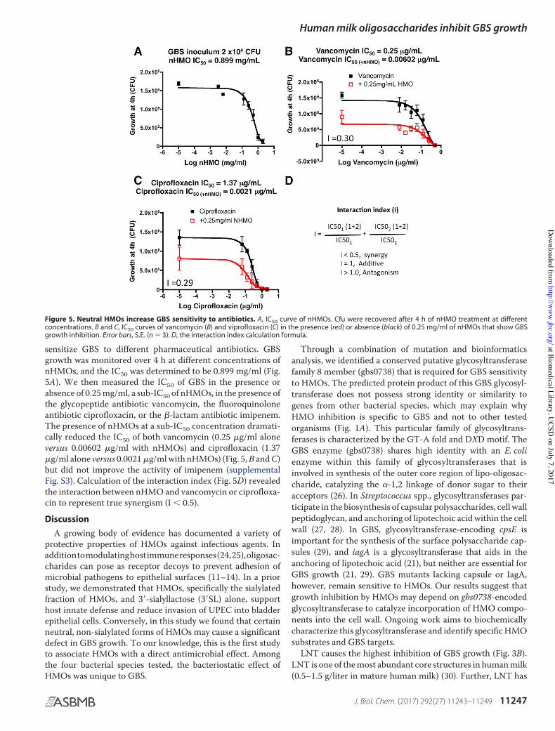
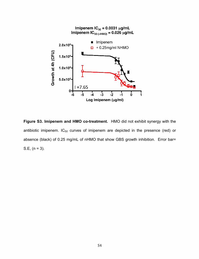
sensitize GBS to different pharmaceutical antibiotics. GBSgrowth was monitored over 4 h at different concentrations ofnHMOs, and the IC50 was determined to be 0.899 mg/ml (Fig.5A). We then measured the IC50 of GBS in the presence orabsence of 0.25 mg/ml, a sub-IC50 of nHMOs, in the presence ofthe glycopeptide antibiotic vancomycin, the fluoroquinoloneantibiotic ciprofloxacin, or the "-lactam antibiotic imipenem.The presence of nHMOs at a sub-IC50 concentration dramati-cally reduced the IC50 of both vancomycin (0.25 !g/ml aloneversus 0.00602 !g/ml with nHMOs) and ciprofloxacin (1.37!g/ml alone versus 0.0021 !g/ml with nHMOs) (Fig. 5, B and C)but did not improve the activity of imipenem (supplementalFig. S3). Calculation of the interaction index (Fig. 5D) revealedthe interaction between nHMO and vancomycin or ciprofloxa-cin to represent true synergism (I # 0.5).Discussion
A growing body of evidence has documented a variety ofprotective properties of HMOs against infectious agents. Inadditiontomodulatinghostimmuneresponses(24,25),oligosac-charides can pose as receptor decoys to prevent adhesion ofmicrobial pathogens to epithelial surfaces (11–14). In a priorstudy, we demonstrated that HMOs, specifically the sialylatedfraction of HMOs, and 3'-sialyllactose (3'SL) alone, supporthost innate defense and reduce invasion of UPEC into bladderepithelial cells. Conversely, in this study we found that certainneutral, non-sialylated forms of HMOs may cause a significantdefect in GBS growth. To our knowledge, this is the first studyto associate HMOs with a direct antimicrobial effect. Amongthe four bacterial species tested, the bacteriostatic effect ofHMOs was unique to GBS.
Through a combination of mutation and bioinformaticsanalysis, we identified a conserved putative glycosyltransferasefamily 8 member (gbs0738) that is required for GBS sensitivityto HMOs. The predicted protein product of this GBS glycosyl-transferase does not possess strong identity or similarity togenes from other bacterial species, which may explain whyHMO inhibition is specific to GBS and not to other testedorganisms (Fig. 1A). This particular family of glycosyltrans-ferases is characterized by the GT-A fold and DXD motif. TheGBS enzyme (gbs0738) shares high identity with an E. colienzyme within this family of glycosyltransferases that isinvolved in synthesis of the outer core region of lipo-oligosac-charide, catalyzing the #-1,2 linkage of donor sugar to theiracceptors (26). In Streptococcus spp., glycosyltransferases par-ticipate in the biosynthesis of capsular polysaccharides, cell wallpeptidoglycan, and anchoring of lipotechoic acid within the cellwall (27, 28). In GBS, glycosyltransferase-encoding cpsE isimportant for the synthesis of the surface polysaccharide cap-sules (29), and iagA is a glycosyltransferase that aids in theanchoring of lipotechoic acid (21), but neither are essential forGBS growth (21, 29). GBS mutants lacking capsule or IagA,however, remain sensitive to HMOs. Our results suggest thatgrowth inhibition by HMOs may depend on gbs0738-encodedglycosyltransferase to catalyze incorporation of HMO compo-nents into the cell wall. Ongoing work aims to biochemicallycharacterize this glycosyltransferase and identify specific HMOsubstrates and GBS targets.
LNT causes the highest inhibition of GBS growth (Fig. 3B).LNT is one of the most abundant core structures in human milk(0.5–1.5 g/liter in mature human milk) (30). Further, LNT has
Figure 5. Neutral HMOs increase GBS sensitivity to antibiotics. A, IC50 curve of nHMOs. Cfu were recovered after 4 h of nHMO treatment at differentconcentrations. B and C, IC50 curves of vancomycin (B) and viprofloxacin (C) in the presence (red) or absence (black) of 0.25 mg/ml of nHMOs that show GBSgrowth inhibition. Error bars, S.E. (n % 3). D, the interaction index calculation formula.
Human milk oligosaccharides inhibit GBS growth
J. Biol. Chem. (2017) 292(27) 11243–11249 11247
at Biomedical Library, U
CSD on July 7, 2017
http://ww
w.jbc.org/
Dow
nloaded from
been identified to block Entamoeba histolytica binding to epi-thelial cell surfaces (12). However, LNT has never been pro-posed to serve any direct antimicrobial function. One intrigu-ing observation is that a slight conformational change of thesingle 3GlcNAc"1 residue of LNT to 4GlcNAc"1 in LNnTnearly abrogates its bacteriostatic effect on GBS (Fig. 3B). Ourresults suggest that there is a strict 3GlcNAC"1 conformationrequirement for maximal GBS inhibition.
HMOs have been found in the plasma (15, 16) and urine (17,18) of breast-fed infants. Also, HMOs appear in the urine ofpregnant women as early as the end of first trimester. Althoughthe exact concentration of HMOs present in these areas is stilluncertain, our results support the notion that lower incidencesof GBS infections in breast-fed infants and already in pregnantwomen could include a contribution from the antibacterialproperties of HMOs. Further, our demonstration of HMO syn-ergism with certain pharmaceutical antibiotics suggests theirpotential utility in adjunctive therapy of GBS infection. Futureanimal studies as well as human cohort studies on humanmother-infant dyads may help identify associations of individ-ual HMOs like LNT with GBS infection risk. It is intriguing toenvision the rational development of novel anti-infective strat-egies based on the natural template of human milk.
Experimental procedures
Bacterial strains, cells, media, and growth conditions
The wild-type GBS strain COH1 (serotype I and I) and itsmutant derivatives HY106 and &iagA (21), A909 (serotype Ia),NCTC10/84 (serotype V), and methicillin-resistant S. aureusstrain TCH1516 were grown overnight in Todd-Hewitt agar orTodd-Hewitt broth (THB). P. aeruginosa strain PA14 andUPEC strain CFT073 (O6:K2:H1, ATCC 700928) were grownovernight in Luria-Bertani agar or broth. All bacteria werepropagated in standing culture to stationary phase at 37 °C in5% CO2 unless stated otherwise.
Human milk oligosaccharide isolations
pHMOs were prepared as described previously (31) andlyophilized for long-term storage. Milk from 36 differentdonors was pooled to account for heterogeneity in HMO com-position between different women. The human milk donationprogram has been reviewed by the Institutional Review Board(IRB) Chair at University of California, San Diego and certifiedas exempt from IRB review under 45 CFR 46.101(b), category 4because subjects cannot be identified and linked to generateddata. Pooled HMOs were separated into aHMO and nHMO byanion exchange chromatography as described previously (31).nHMOs were further separated by size using size exclusionchromatography. pHMOs were disialylated by incubation withneuraminidase from Vibrio cholerae. HMO composition wasanalyzed by high-performance liquid chromatography andmass spectrometry.
Sources of galacto-oligosaccharides and purified glycans
Galacto-oligosaccharides were generously provided by Frie-sland Campina Domo (the Netherlands). Purified glycans werepurchased from ELICITYL OligoTech!, which includes LNT,
LNnT, LNnH, LNFPI, LNDFHII, LnNO, LNnDFH, LNnFPV,and LNFPV.
GBS growth and susceptibility test
Overnight GBS growth was back-diluted to A600 % 0.01 inserum-free RPMI 1640 (SF-RPMI) (Life Technologies) or sup-plemented with pooled or fractions of HMOs. To measuregrowth, bacteria were grown in 100 !l of medium at 37 °C. At600 nm, absorbance was recorded at 30-min interval using theBioScreen instrument (Growth Curves USA). To detect growthin cfu, bacteria were grown in 100 !l of medium at 37 °C inhumidified air with 5% CO2 over 2 or 4 h. Recombinant pro-teinase K (Roche) was used at 0.1 mg/ml.
GBS COH-1 transposon library
The transposon library was constructed as described previ-ously (21). To screen for mutants resistant to HMOs, we nor-malized overnight cultures of transposon mutants to A600 of0.1. Approximately 10 !l of each mutant was cultured in 100 !lof SF-RPMI supplemented with 5 mg/ml of pooled HMOs.Growth was monitored over an 8-h period at 37 °C by measur-ing optical density at 600 nm at a 30-min interval using a Bio-screen C MBR system.
GBS COH-1 !0738 construct
To generate a targeted knockout, we cloned the gbs0738gene to the temperature-sensitive vector PHY304. Briefly,gbs0738 was PCR-amplified with primers XhoI-gbs0738F (5'-CGATCTCGAGTGCTCAGGCACCTACAACTG-3') ( Hin-dIII-gbs0738R (5'-CAGTAAGCTTAGCAGGCAAGTTCAT-CAAGAG-3') to generate a 300-bp amplicon. The purifiedPCR amplicon was digested with XhoI and HindIII and ligatedinto previously digested pHY304. The construct was clonedinto E. coli DH5# and isolated by mini-prep. Approximately 1!g of PHY304-gbs0738 DNA was transformed into electro-competent GBS at 1550 V. The transformed bacteria wereallowed to recover in 500 !l of THB with 0.25 M sucrose at 30 °Cfor 2 h while shaking at 220 RPM. Bacteria were grown over-night on Todd-Hewitt agar ( 2 !g/ml erythromycin at 30 °C.After 2–3 days, colonies were selected and inoculated into 5 mlof THB ( 5 !g/ml erythromycin at 30 °C overnight. The nextday, cultures were prepared in duplicate in THB ( erythromy-cin at 30 °C and 37 °C to select for bacteria with the targetingvector incorporated into the chromosome. Resultant single col-onies from the 37 °C culture were isolated and confirmed forsingle crossover mutation by PCR using primers M13F (5'-GTTTTCCCAGTCACGAC-3') and gbs0738R2 (5'-ACACG-CTCCTCCTTTGATATT-3'). Wild-type GBS had an ex-pected 1.67 kb PCR product, whereas the mutant had anexpected 6.5-kbp PCR product.
Statistical analysis
All experiments were performed in triplicate or quadrupli-cate and repeated in at least two independent experiments.Error bars represent S.E. (n ! 3) from multiple independentexperiments. Statistical analysis was performed using Student’sunpaired two-tailed t test or one-way ANOVA for multiplecomparisons (GraphPad Prism version 5.03). *, p # 0.05; **, p #
Human milk oligosaccharides inhibit GBS growth
11248 J. Biol. Chem. (2017) 292(27) 11243–11249
at Biomedical Library, U
CSD on July 7, 2017
http://ww
w.jbc.org/
Dow
nloaded from
0.01; and ***, p # 0.001 represent statistical significance. p !0.05 represents non-significance. IC50 was calculated usingnon-linear regression curve fit using the equation log (inhibi-tor) versus response-variable slope. Error bars represent S.E.(n % 3).
Author contributions—A. E. L. designed and performed the experi-ments, analyzed the data, prepared the figures, and co-wrote themanuscript. C. A. A., A. S., M. H., K. G., and T. E. performed theexperiments. A. R. P. and G. J. B. provided the desialylated HMOs.K. S. D. provided the GBS transposon library. A. L. L. and K. G. par-ticipated in data analysis. L. B. and V. N. designed the experiments,participated in data analysis, and co-wrote the manuscript.
Acknowledgments—We thank Samira Dahesh for expertise regardingGBS cloning and Federico C. Beasley for helpful discussions regardingdata interpretation and critical review of the manuscript.
References1. Edwards, M. S. (2006) Issues of antimicrobial resistance in group B strep-
tococcus in the era of intrapartum antibiotic prophylaxis. Semin. Pediatr.Infect. Dis. 17, 149 –152
2. Heath, P. T., and Schuchat, A. (2007) Perinatal group B streptococcaldisease. Best. Pract. Res. Clin. Obstet. Gynaecol. 21, 411– 424
3. Thigpen, M. C., Whitney, C. G., Messonnier, N. E., Zell, E. R., Lynfield, R.,Hadler, J. L., Harrison, L. H., Farley, M. M., Reingold, A., Bennett, N. M.,Craig, A. S., Schaffner, W., Thomas, A., Lewis, M. M., Scallan, E., et al.(2011) Bacterial meningitis in the United States, 1998 –2007. N. Engl.J. Med. 364, 2016 –2025
4. Phares, C. R., Lynfield, R., Farley, M. M., Mohle-Boetani, J., Harrison, L. H.,Petit, S., Craig, A. S., Schaffner, W., Zansky, S. M., Gershman, K., Stefonek,K. R., Albanese, B. A., Zell, E. R., Schuchat, A., Schrag, S. J., and ActiveBacterial Core Surveillance/Emerging Infections Program Network(2008) Epidemiology of invasive group B streptococcal disease in theUnited States, 1999 –2005. JAMA 299, 2056 –2065
5. Bedford, H., de Louvois, J., Halket, S., Peckham, C., Hurley, R., and Harvey,D. (2001) Meningitis in infancy in England and Wales: follow up at age 5years. BMJ 323, 533–536
6. Edmond, K. M., Kortsalioudaki, C., Scott, S., Schrag, S. J., Zaidi, A. K.,Cousens, S., and Heath, P. T. (2012) Group B streptococcal disease ininfants aged younger than 3 months: systematic review and meta-analysis.Lancet 379, 547–556
7. Baltimore, R. S. (2007) Consequences of prophylaxis for group B strepto-coccal infections of the neonate. Semin. Perinatol. 31, 33–38
8. Castor, M. L., Whitney, C. G., Como-Sabetti, K., Facklam, R. R., Ferrieri,P., Bartkus, J. M., Juni, B. A., Cieslak, P. R., Farley, M. M., Dumas, N. B.,Schrag, S. J., and Lynfield, R. (2008) Antibiotic resistance patterns in inva-sive group B streptococcal isolates. Infect Dis. Obstet. Gynecol. 2008,727505
9. Bode, L., and Jantscher-Krenn, E. (2012) Structure-function relationshipsof human milk oligosaccharides. Adv. Nutr. 3, 383S-391S
10. Bode, L. (2012) Human milk oligosaccharides: every baby needs a sugarmama. Glycobiology 22, 1147–1162
11. Martín-Sosa, S., Martín, M. J., and Hueso, P. (2002) The sialylated fractionof milk oligosaccharides is partially responsible for binding to enterotoxi-genic and uropathogenic Escherichia coli human strains. J. Nutr. 132,3067–3072
12. Jantscher-Krenn, E., Lauwaet, T., Bliss, L. A., Reed, S. L., Gillin, F. D., andBode, L. (2012) Human milk oligosaccharides reduce Entamoeba histo-lytica attachment and cytotoxicity in vitro. Br. J. Nutr. 108, 1839 –1846
13. Newburg, D. S., Pickering, L. K., McCluer, R. H., and Cleary, T. G. (1990)Fucosylated oligosaccharides of human milk protect suckling mice fromheat-stabile enterotoxin of Escherichia coli. J. Infect. Dis. 162, 1075–1080
14. Newburg, D. S. (2005) Innate immunity and human milk. J. Nutr. 135,1308 –1312
15. Ruhaak, L. R., Stroble, C., Underwood, M. A., and Lebrilla, C. B. (2014)Detection of milk oligosaccharides in plasma of infants. Anal. Bioanal.Chem. 406, 5775–5784
16. Goehring, K. C., Kennedy, A. D., Prieto, P. A., and Buck, R. H. (2014) Directevidence for the presence of human milk oligosaccharides in the circula-tion of breastfed infants. PLoS ONE 9, e101692
17. Rudloff, S., Obermeier, S., Borsch, C., Pohlentz, G., Hartmann, R.,Brösicke, H., Lentze, M. J., and Kunz, C. (2006) Incorporation of orallyapplied 13C-galactose into milk lactose and oligosaccharides. Glycobiology16, 477– 487
18. Dotz, V., Rudloff, S., Meyer, C., Lochnit, G., and Kunz, C. (2015) Metabolicfate of neutral human milk oligosaccharides in exclusively breast-fed in-fants. Mol. Nutr. Food Res. 59, 355–364
19. Hallgren, P., and Lundblad, A. (1977) Structural analysis of nine oligosac-charides isolated from the urine of a blood group O, nonsecretor, womanduring pregnancy and lactation. J. Biol. Chem. 252, 1014 –1022
20. Lin, A. E., Autran, C. A., Espanola, S. D., Bode, L., and Nizet, V. (2014)Human milk oligosaccharides protect bladder epithelial cells against uro-pathogenic Escherichia coli invasion and cytotoxicity. J. Infect. Dis. 209,389 –398
21. Doran, K. S., Engelson, E. J., Khosravi, A., Maisey, H. C., Fedtke, I., Equils,O., Michelsen, K. S., Arditi, M., Peschel, A., and Nizet, V. (2005) Blood-brain barrier invasion by group B Streptococcus depends upon propercell-surface anchoring of lipoteichoic acid. J. Clin. Invest. 115, 2499 –2507
22. Parker, C. T., Pradel, E., and Schnaitman, C. A. (1992) Identification andsequences of the lipopolysaccharide core biosynthetic genes rfaQ, rfaP,and rfaG of Escherichia coli K-12. J. Bacteriol. 174, 930 –934
23. Yim, H. H., Nittayarin, A., and Rubens, C. E. (1997) Analysis of the capsulesynthesis locus, a virulence factor in group B streptococci. Adv. Exp. Med.Biol. 418, 995–997
24. He, Y., Liu, S., Kling, D. E., Leone, S., Lawlor, N. T., Huang, Y., Feinberg,S. B., Hill, D. R., and Newburg, D. S. (2016) The human milk oligosaccha-ride 2'-fucosyllactose modulates CD14 expression in human enterocytes,thereby attenuating LPS-induced inflammation. Gut 65, 33– 46
25. Li, M., Monaco, M. H., Wang, M., Comstock, S. S., Kuhlenschmidt, T. B.,Fahey, G. C., Jr., Miller, M. J., Kuhlenschmidt, M. S., and Donovan, S. M.(2014) Human milk oligosaccharides shorten rotavirus-induced diarrheaand modulate piglet mucosal immunity and colonic microbiota. ISME J. 8,1609 –1620
26. Leipold, M. D., Vinogradov, E., and Whitfield, C. (2007) Glycosyltrans-ferases involved in biosynthesis of the outer core region of Escherichiacoli lipopolysaccharides exhibit broader substrate specificities than ispredicted from lipopolysaccharide structures. J. Biol. Chem. 282,26786 –26792
27. Miyake, K., and Iijima, S. (2004) Bacterial capsular polysaccharide andsugar transferases. Adv. Biochem. Eng. Biotechnol. 90, 89 –111
28. Shainheit, M. G., Valentino, M. D., Gilmore, M. S., and Camilli, A. (2015)Mutations in pneumococcal cpsE generated via in vitro serial passagingreveal a potential mechanism of reduced encapsulation utilized by a con-junctival isolate. J. Bacteriol. 197, 1781–1791
29. Cieslewicz, M. J., Kasper, D. L., Wang, Y., and Wessels, M. R. (2001) Func-tional analysis in type Ia group B Streptococcus of a cluster of genes in-volved in extracellular polysaccharide production by diverse species ofstreptococci. J. Biol. Chem. 276, 139 –146
30. Pfenninger, A., Karas, M., Finke, B., and Stahl, B. (2002) Structural analysisof underivatized neutral human milk oligosaccharides in the negative ionmode by nano-electrospray MS(n) (part 2: application to isomeric mix-tures). J. Am. Soc. Mass Spectrom. 13, 1341–1348
31. Jantscher-Krenn, E., Zherebtsov, M., Nissan, C., Goth, K., Guner, Y. S.,Naidu, N., Choudhury, B., Grishin, A. V., Ford, H. R., and Bode, L. (2012)The human milk oligosaccharide disialyllacto-N-tetraose prevents necro-tising enterocolitis in neonatal rats. Gut 61, 1417–1425
Human milk oligosaccharides inhibit GBS growth
J. Biol. Chem. (2017) 292(27) 11243–11249 11249
at Biomedical Library, U
CSD on July 7, 2017
http://ww
w.jbc.org/
Dow
nloaded from
S1
SUPPLEMENTAL MATERIAL
Human Milk Oligosaccharides Inhibit Growth of Group B Streptococcus
Ann E. Lin1, Chloe A. Autran2,3, Alexandra Szyszka2,3, Tamara Escajadillo1, Mia Huang4,
Kamil Godula4, Anthony R. Prudden7, Geert-Jan Boons7, Amanda L. Lewis8,
Kelly S. Doran1,9, Victor Nizet1,5,6 and Lars Bode2,3,6,10*
Figure S1 GBS growth in pHMO with or without proteinase K ........................... Page S2
Figure S2 gbs0738 AA sequence alignment. ..................................................... Page S3
Figure S3 Imipenem and HMO co-treatment ...................................................... Page S4
S2
Figure S1. GBS growth in pHMO with or without proteinase K. GBS serotype III
strain COH1 was incubated in SF-RPMI alone (red), with 5 mg/mL pHMO (black), or
with addition of 0.1 mg/mL of Proteinase K (gray) over 6 h.
S3
Figure S2. gbs0738 AA sequence alignment
S4
Figure S3. Imipenem and HMO co-treatment. HMO did not exhibit synergy with the
antibiotic imipenem. IC50 curves of imipenem are depicted in the presence (red) or
absence (black) of 0.25 mg/mL of nHMO that show GBS growth inhibition. Error bar=
S.E, (n = 3).