lymphangiomyomatosis presentation dr. med. i. nenadic
DESCRIPTION
TRANSCRIPT
Lymphangioleiomyomatosis of the lung
Dr. med. I. Nenadić
Prof. Dr. med. S. Störkel
HELIOS Klinikum Wuppertal
Institut für Pathologie
1
History: 34 yo female patient, suspicion of
lymphangioleiomyomatosis
Macroscopy: Lung resection specimen of segment V of left
upper lobe of the lung 4 x 4 x 2 cm in size
Histology: H 09713/11
2
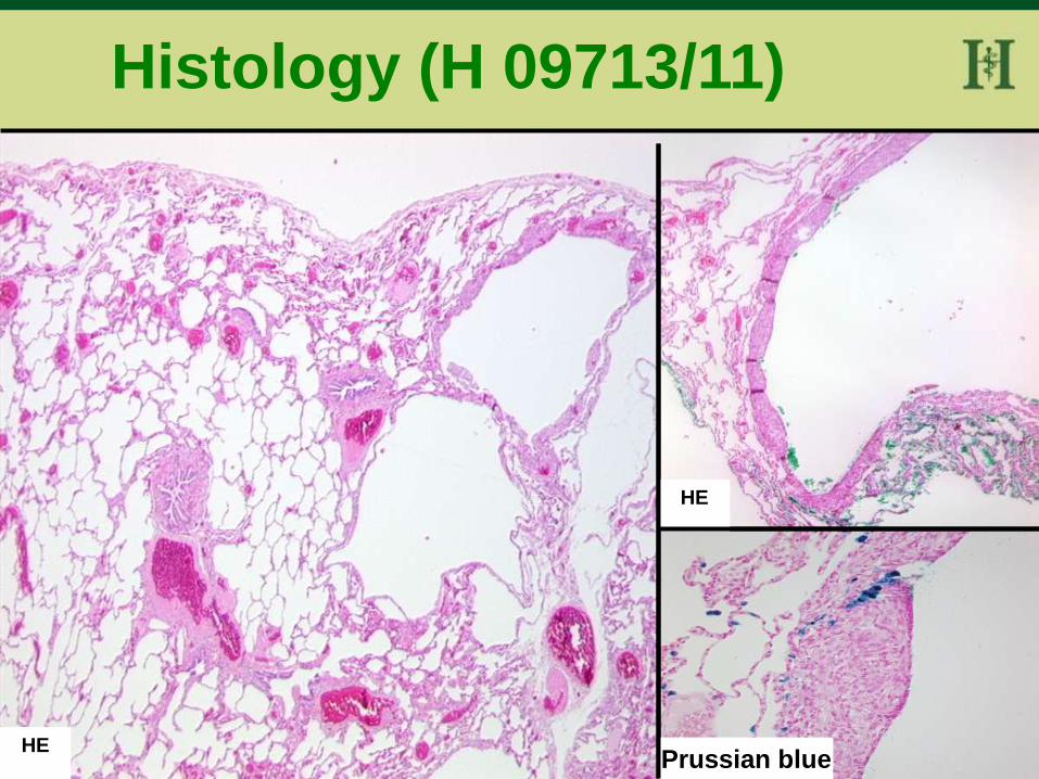
Histology (H 09713/11)
HE
HE
Prussian blue
3
Berliner Blau Färbung HE HE
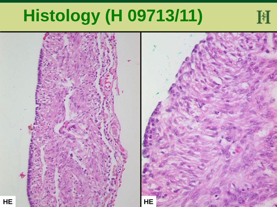
Histology (H 09713/11)
4
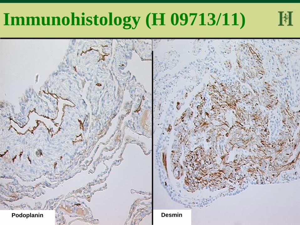
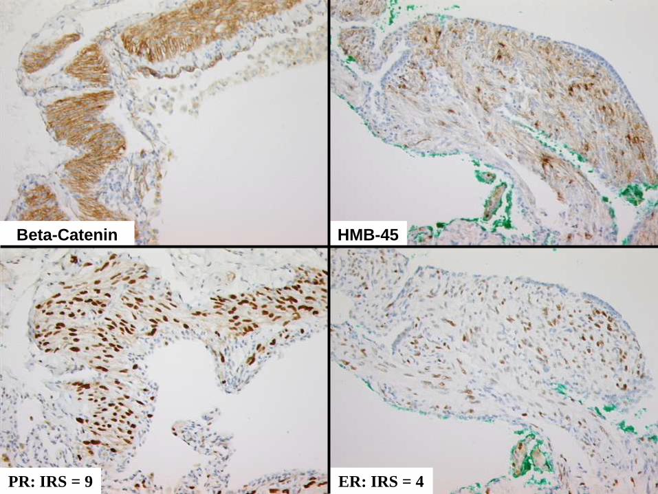
Immunohistology (H 09713/11)
Podoplanin Desmin
5
Kolo
n
Kolon
Beta-Catenin HMB-45
PR: IRS = 9 ER: IRS = 4
6
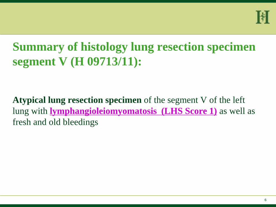
Summary of histology lung resection specimen
segment V (H 09713/11):
Atypical lung resection specimen of the segment V of the left
lung with lymphangioleiomyomatosis (LHS Score 1) as well as
fresh and old bleedings
7
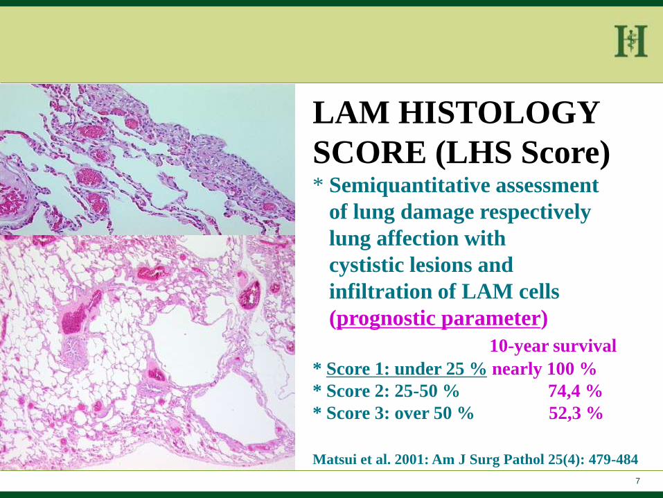
LAM HISTOLOGY
SCORE (LHS Score) * Semiquantitative assessment
of lung damage respectively
lung affection with
cystistic lesions and
infiltration of LAM cells
(prognostic parameter)
10-year survival * Score 1: under 25 % nearly 100 %
* Score 2: 25-50 % 74,4 %
* Score 3: over 50 % 52,3 %
Matsui et al. 2001: Am J Surg Pathol 25(4): 479-484
8
LYMPHANGIOLEIOMYOMATOSIS * affects nearly exclusively women in reproductive age
* Incidence 1-2,6 cases / 1000000 women (often misdiagnosed !)
* Described in the 60´s in the literature for the first time
Cornog JL et al. Cancer 1966;19:1909-1930
* Outcome variable, median survival time of 8-10 years after
diagnosis
9
* Monoclonal proliferation of immature respectively abnormal smooth
muscle cells in the lungs, in the wall of lymphatic vessels (partially
also in bronchioles) in and lymp nodes in the thorax and
retroperitoneum) with progressive cystic destruction of the lungs
* Pathognomonic HMB-45 expression (melanocytic marker)
LYMPHANGIOLEIOMYOMATOSIS
10
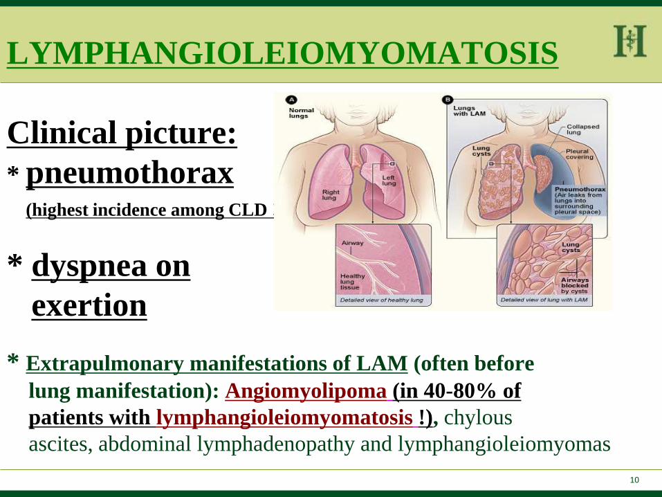
LYMPHANGIOLEIOMYOMATOSIS
Clinical picture:
* pneumothorax (highest incidence among CLD !)
* dyspnea on
exertion
* Extrapulmonary manifestations of LAM (often before
lung manifestation): Angiomyolipoma (in 40-80% of
patients with lymphangioleiomyomatosis !), chylous
ascites, abdominal lymphadenopathy and lymphangioleiomyomas
11
PEComa
perivascular epitheloid cell tumour • Mesenchymal tumor with relatedness to smooth
muscle cells and melanocytes (positivity for melanocytic and myogenic markers), origin from vessel walls and mainly benign course
• Angiomyolipoma
• Lymphangioleiomyomatosis
• High prevalence of angiomyolipoma in lymphangiomyomatosis!
• Clear cell sugar tumour of the lung
12
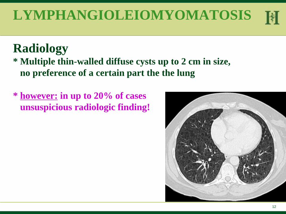
LYMPHANGIOLEIOMYOMATOSIS
Radiology * Multiple thin-walled diffuse cysts up to 2 cm in size,
no preference of a certain part the the lung
* however: in up to 20% of cases
unsuspicious radiologic finding!
13
LYMPHANGIOLEIOMYOMATOSIS
Therapy: no effective treatment options! Harari S et al. 2011; Eur Respir Rev 20: 119, 34-44
* Estrogen blocker (not proofed !)
* Progesteron preparation (consider in single cases with
rapid progression)
* Ultima ratio treatment: lung transplantation (better
survival rate of LAM-patients than in
other patient groups after lung transplantation)
14
LYMPHANGIOLEIOMYOMATOSIS
• Sporadic form (mainly with more pronounced clinical
symptoms)
• In tuberous sclerosis (30-40 % of patients)
• Associated with mutations of genes of tuberous sclerosis
TSC1 or TSC2 with consecutive proliferation of immature
respectively abnormal muscle cells
15
Genes of tuberous sclerosis
TSC1 and TSC2 tumor suppressor gene
• Hamartin: Protein product of TSC1 gene (reorganization of actin cytoskeleton)
• Tuberin: Protein product of TSC2 gene (negative regulator of cell cycle progression)
• Hamartin-tuberin complex: negative regulator of mTOR
• Loss of TSC function results in mTOR activation
• Hypothesis: Rapamycin (mTOR inhibitor) could influence LAM progression ?
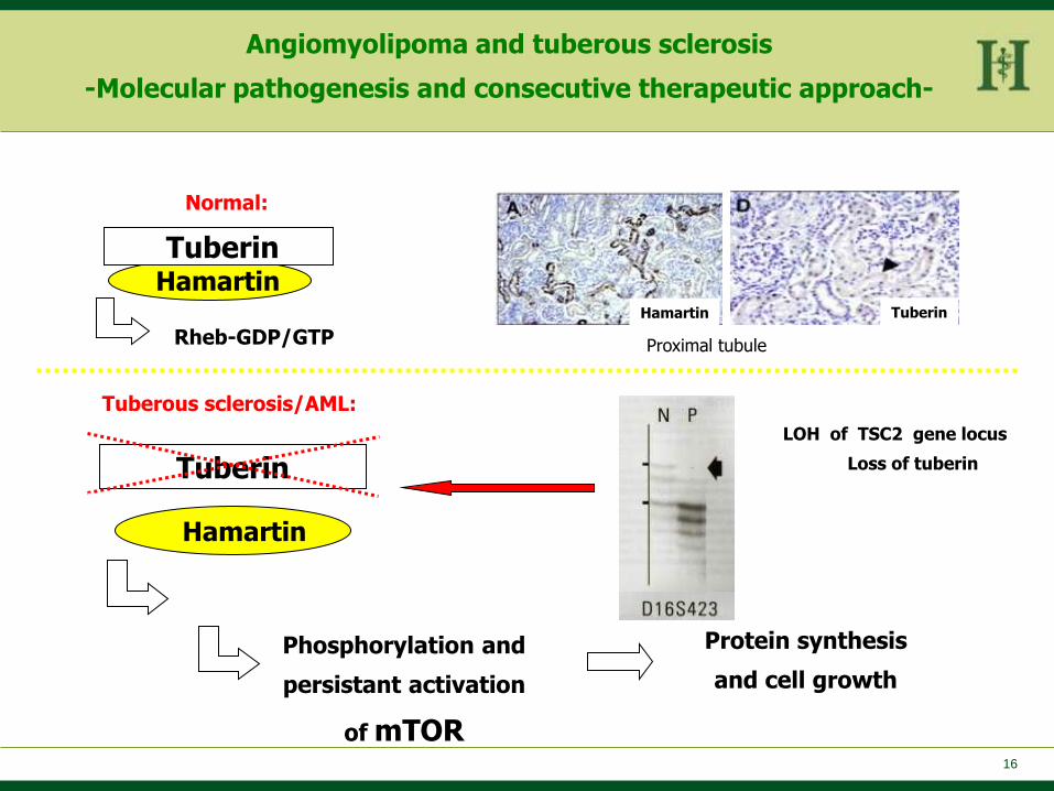
16
Rheb-GTP
Hamartin
Tuberin
Tuberous sclerosis/AML:
Phosphorylation and
persistant activation
of mTOR
Protein synthesis
and cell growth
Hamartin
Tuberin
Normal:
Rheb-GDP/GTP
Hamartin and tuberin expression in renal cortex
Tuberin Hamartin
Proximal tubule
LOH of TSC2 gene locus
Loss of tuberin
Angiomyolipoma and tuberous sclerosis
-Molecular pathogenesis and consecutive therapeutic approach-
TSC2:
TSC1:
TSC2:
TSC1:
17
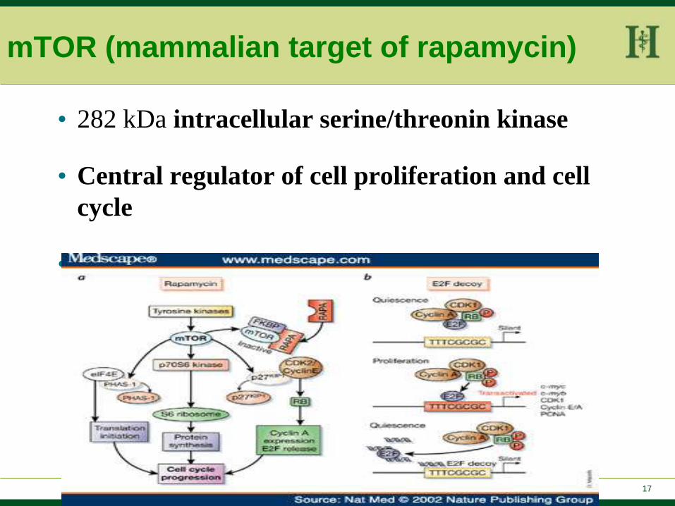
mTOR (mammalian target of rapamycin)
• 282 kDa intracellular serine/threonin kinase
• Central regulator of cell proliferation and cell
cycle
• mTOR activation described in PEComas
19
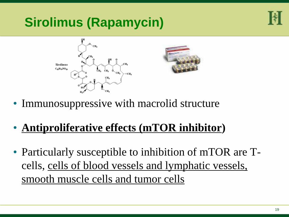
Sirolimus (Rapamycin)
• Immunosuppressive with macrolid structure
• Antiproliferative effects (mTOR inhibitor)
• Particularly susceptible to inhibition of mTOR are T-
cells, cells of blood vessels and lymphatic vessels,
smooth muscle cells and tumor cells
20
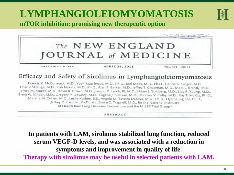
LYMPHANGIOLEIOMYOMATOSIS mTOR inhibition: promising new therapeutic option
In patients with LAM, sirolimus stabilized lung function, reduced
serum VEGF-D levels, and was associated with a reduction in
symptoms and improvement in quality of life.
Therapy with sirolimus may be useful in selected patients with LAM.
21
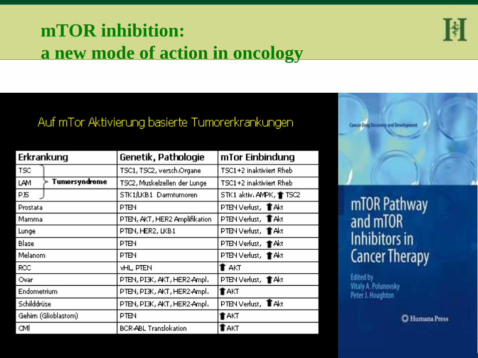
mTOR inhibition:
a new mode of action in oncology
22
SUMMARY
Lymphangiomyomatosis: rare differential diagnosis of
cystic lung diseases
In young women with pneumothorax, angiomyolipoma of
the kidney or tuberous sclerosis LAM should always be
considered as a differential diagnosis
Exact and early diagnosis and differentiation from other
(diffuse) cystistic lung diseases is important due to
different therapeutic approaches

23 Titel Präsentation, Thema, Verfasser, Datum HELIOS Standort
www.helios-kliniken.de
Thank you for your attention! HELIOS Klinikum Wuppertal
Instutut für Patthologie

24 Titel Präsentation, Thema, Verfasser, Datum HELIOS Standort
www.helios-kliniken.de
Thank you for your attention! HELIOS Klinikum Wuppertal
Institut für Pathologie