poster shubham chattopadhyay
TRANSCRIPT
DBP
DBPMYBBP1A
!"#$!"#%!"#&!"#'!"#(!"#)!"#*!"#+!"#,!"#
$!!"#
$%# %'# &)#
!"#$%
"&'(")(**+"
,-./+"%-+0"123(45-2"
678("19"
678("111"
678("11"
678("1"
Investigating the Regulation of Nucleolar Proteins by Adenovirus Infection Shubham Chattopadhyay, Cipriano Zuluaga, Emigdio D. Reyes1, Daniel K. Bricker1, Matthew D. Weitzman1
1Department of Pathology and Laboratory Medicine, Children’s Hospital of Philadelphia and University of Pennsylvania Perelman School of Medicine
Abstract
Adenovirus Alters Subcellular Structure
NucleusCytoplasm
Nucleolar proteins Cellular
DNA
Nuclear Proteins Nucleus
Cytoplasm
VRC VRC
Viral Protein
iPONDMS
V H
Nucleolar Proteins enriched on vDNA
Enriched Proteins
What role do nucleolar factors play Ad infection?
Adenovirus (Ad) has been utilized as a model system to study key cellular processes such as replication, transcription, and apoptosis. An obligate intracellular parasite, Ad must hijack host cellular machinery and manipulate host factors in order to replicate, assemble, and disperse viral progeny. Ad forms viral replication centers (VRCs), the sites of viral replication, within nuclei of infected cells. Our lab is using proteomic approaches to identify proteins recruited to replicating viral DNA to potentially identify novel host proteins involved in viral replication. These studies revealed many nucleolar proteins that associated with Ad nascent viral DNA. Here, we expanded on this proteomic data by defining the expression level, modification status, and subcellular localization of a subset of these nucleolar proteins during adenovirus infection. We employed western blot analysis and immunofluorescence to compare uninfected (Host) cells with cells infected with wild-type adenovirus serotype 5 (Ad5). We found that nucle-olar proteins accumulated with a marker of VRCs, formed foci surrounding VRCs, or were dis-persed from the nucleolus to the nucleoplasm in infected cells. We also determined that sever-al non-nucleolar proteins were affected by adenovirus infection. Taken together, these results suggest that Ad impacts nucleolar biology during infection. We hypothesize that Ad modulates nucleolar proteins to promote aspects of the viral life cycle, such as viral transcription or pro-cessing of viral RNAs. To characterize further the role of nucleolar factors during Ad infection, we propose to employ siRNA knockdown of selective nucleolar proteins in Ad5-infected cells.
• Where do nucleolar proteins localize during viral replication? • To what extent is overall abundance of nucleolar proteins affected by Ad infection?
Nucleolar Transcriptional Regulators Form Foci around Ad VRCs
Experimental Approach
Viral InfectionAbundance Localization
B. Coilin forms distinct foci around Ad5 VRCs
A. UBF and Dyskerin affected by Ad5 infection
Coi
linD
BP
+ m
erge
rRNA Maturation Proteins Disperse from Nucleolar Bodies upon Ad Infection
A. DDX21 abundance level impacted by Ad5 infection
C. DDX21 displays pan-nuclear dispersion during infection
B. DDX21 diffuses to nucleoplasm from nucleoli
DBP marks viral DNA in Ad5 infected A549 cells. DDX21 localiza-tion pattern above. Cells grouped into four localization patterns & counted at each infection point, below. N = 45, 96, 97.
DD
X21D
BP
+ m
erge
rRNA Processing Proteins Accumulate at Ad VRCs A. NOLC1 is altered by Ad5
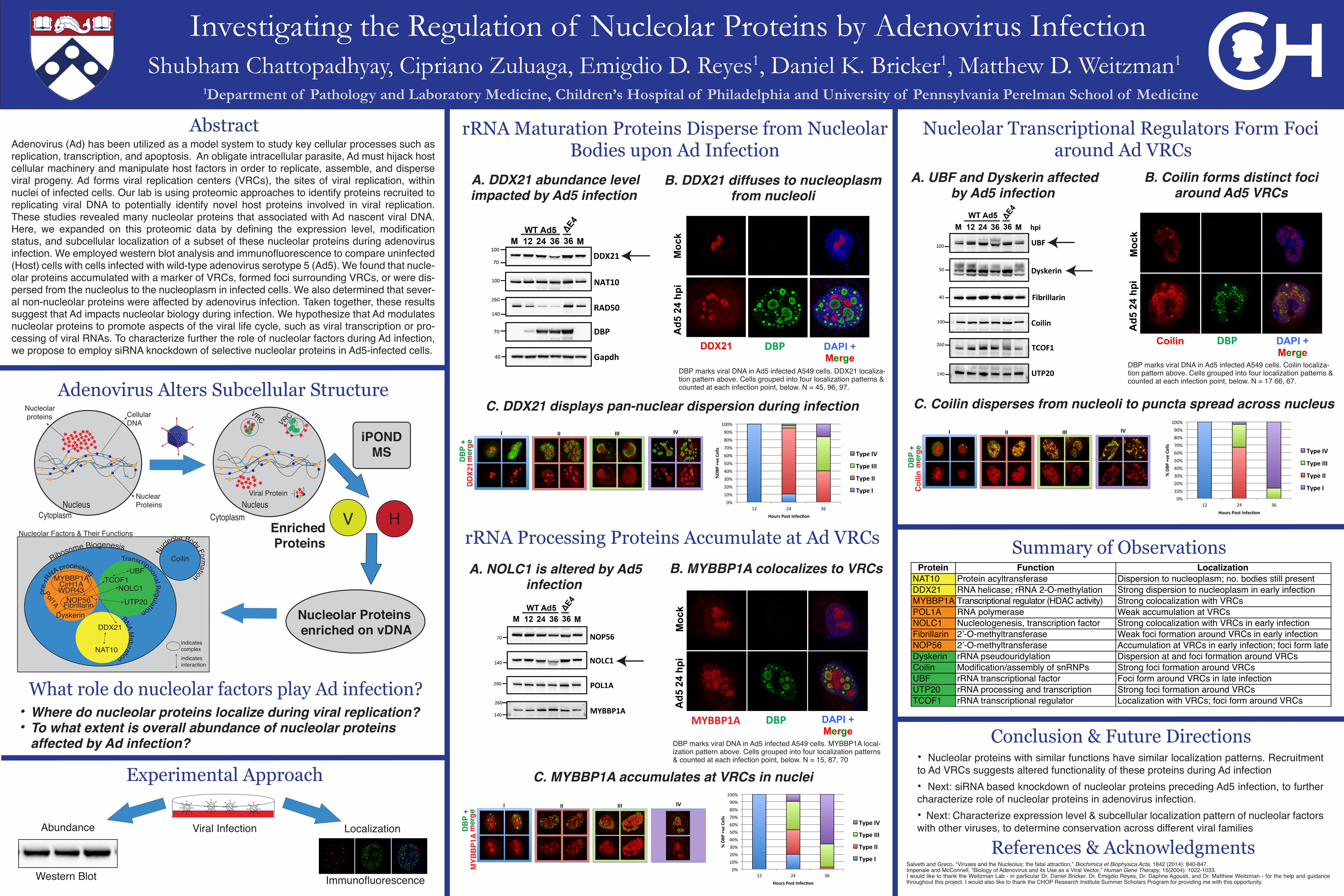
infection B. MYBBP1A colocalizes to VRCs
DBP marks viral DNA in Ad5 infected A549 cells. Coilin localiza-tion pattern above. Cells grouped into four localization patterns & counted at each infection point, below. N = 17 66, 67.
DBP marks viral DNA in Ad5 infected A549 cells. MYBBP1A local-ization pattern above. Cells grouped into four localization patterns & counted at each infection point, below. N = 15, 87, 70
C. MYBBP1A accumulates at VRCs in nuclei
MYB
BP1
AD
BP
+ m
erge
Summary of Observations
References & Acknowledgments
Nu
cleolar Body Formation
CoilinRibosome Biogenesis
RNA Maturation
pre-
rR
NA processingTranscriptional Regulation
NOP56Fibrillarin
MYBBP1ACirH1AWDR43
NAT10
DDX21
TCOF1
UTP20
Dyskerin
NOLC1
UBF
indicatesinteraction
indicatescomplex
Pol1A
Nucleolar Factors & Their Functions
!"#$!"#%!"#&!"#'!"#(!"#)!"#*!"#+!"#,!"#$!!"#
$%# %'# &)#
!"#$%
"&'(")(**+"
,-./+"%-+0"123(45-2"
678("19"
678("111"
678("11"
678("1"
!"#
$!"#
%!"#
&!"#
'!"#
(!"#
)!"#
*!"#
+!"#
,!"#
$!!"#
$%# %'# &)#
!"#
$%&'(%)(
**+%
,-./+%$-+0%123(45-2%
678(%19%
678(%111%
678(%11%
678(%1%
Salvetti and Greco, “Viruses and the Nucleolus: the fatal attraction,” Biochimica et Biophysica Acta, 1842 (2014): 840-847. Imperiale and McConnell, “Biology of Adenovirus and its Use as a Viral Vector,” Human Gene Therapy, 15(2004): 1022-1033. I would like to thank the Weitzman Lab - in particular Dr. Daniel Bricker, Dr. Emigdio Reyes, Dr. Daphne Agousti, and Dr. Matthew Weitzman - for the help and guidance throughout this project. I would also like to thank the CHOP Research Institute Summer Scholars Program for providing me with this opportunity.
DBP
Western Blot Immunofluorescence
• Nucleolar proteins with similar functions have similar localization patterns. Recruitment to Ad VRCs suggests altered functionality of these proteins during Ad infection• Next: siRNA based knockdown of nucleolar proteins preceding Ad5 infection, to further characterize role of nucleolar proteins in adenovirus infection. • Next: Characterize expression level & subcellular localization pattern of nucleolar factors with other viruses, to determine conservation across different viral families
Conclusion & Future Directions
Protein Function LocalizationNAT10 Protein acyltransferase Dispersion to nucleoplasm; no. bodies still presentDDX21 RNA helicase; rRNA 2-O-methylation Strong dispersion to nucleoplasm in early infectionMYBBP1A Transcriptional regulator (HDAC activity) Strong colocalization with VRCs POL1A RNA polymerase Weak accumulation at VRCs NOLC1 Nucleologenesis, transcription factor Strong colocalization with VRCs in early infectionFibrillarin 2’-O-methyltransferase Weak foci formation around VRCs in early infectionNOP56 2’-O-methyltransferase Accumulation at VRCs in early infection; foci form lateDyskerin rRNA pseudouridylation Dispersion at and foci formation around VRCsCoilin Modification/assembly of snRNPs Strong foci formation around VRCs UBF rRNA transcriptional factor Foci form around VRCs in late infectionUTP20 rRNA processing and transcription Strong foci formation around VRCs TCOF1 rRNA transcriptional regulator Localization with VRCs; foci form around VRCs
C. Coilin disperses from nucleoli to puncta spread across nucleus