hemangioma kapiler
TRANSCRIPT
672Original Article
Retinal capillary hemangiomas are benign vascu-lar tumors in the retina or optic nerve head.
They are orange-red spherical tumors fed by a dilat-ed, tortuous retinal artery and drained by anengorged vein.(1) The tumor can present as beingeither endophytic or exophytic.(2) Retinal capillaryhemangiomas occurring as isolated tumors have also
been called angiomatosis retinae, retinal angiomato-sis, retinal hemangioblastoma, von Hippel's disease,and von Hippel tumor. Those occurring with tumorsin other systems, especially central nervous systemhemangioblastomas and renal cell carcinomas, werenamed von Hippel-Lindau (VHL) disease.(3,4) VHLdisease is a hereditary familial multiple-system can-
Retinal Capillary Hemangiomas: Clinical Manifestations and Visual Prognosis
Ming-Tse Kuo, MD; Hsi-Kung Kou, MD; Min-Lun Kao, MD; Ming-Hsiung Tsai, MD;Yung-Jen Chen, MD; Sue-Ann Lin, MD
Background: To describe the clinical features, visual outcomes, and therapeutic complica-tions of patients with retinal capillary hemangiomas.
Methods: A retrospective, non-comparative, observational case study of patients diag-nosed with retinal capillary hemangiomas was conducted. Twelve patients(13 eyes) at Chang Gung Memorial Hospital of Kaohsiung from July 1987 toJune 2001 were reviewed. Pre- and post-treatment visual acuity and ocularcomplications are described.
Results: One patient had bilateral and another had unilateral juxtapapillary heman-giomas. All of the other 10 patients were diagnosed with peripheral retinalcapillary hemangiomas. More patients had retinal capillary hemangiomaslocated in the temporal peripheral retina and all had endophytic growth pat-terns. No patient met the diagnostic criteria of von Hippel-Lindau disease.Visual acuity levels of peripheral retinal hemangiomas without exudativeretinal detachment often remained the same after focal laser treatment. Twopatients received vitreoretinal surgery. Patients with juxtapapillary heman-giomas had variable visual outcomes and visual field defects during follow-up.
Conclusion: Early diagnosis of capillary hemangiomas in the retinal periphery and treat-ment by focal laser produced good visual outcomes. If untreated, the tumorsmay eventually be complicated with exudative retinal detachment and have aworse visual prognosis even with vitreoretinal surgery.(Chang Gung Med J 2002;25:672-82)
Key words: retinal capillary hemangioma, von Hippel-Lindau (VHL) disease, exudative retinaldetachment.
From the Department of Ophthalmology, Chang Gung Memorial Hospital, Kaohsiung.Received: Apr. 19, 2002; Accepted: Jul. 25, 2002Address for reprints: Dr. Hsi-Kung Kou, Department of Ophthalmology, Chang Gung Memorial Hospital, Kaohsiung, 123 Ta-PeiRoad, Niaosung 833, Kaohsiung, Taiwan, R.O.C. Tel.: 886-7-7317123 ext. 2814; Fax: 886-7-7317123 ext. 2830; E-mail:[email protected]
Chang Gung Med J Vol. 25 No. 10October 2002
Ming-Tse Kuo, et alRetinal capillary hemangiomas
673
cer syndrome. The cardinal features include retinalcapillary hemangiomas, central nervous systemhemangioblastomas, renal cell carcinomas,pheochromocytomas, and pancreatic, renal, and epi-didymal cysts.(5) Retinal capillary hemangiomas arethe most common presenting feature and often thefirst manifestation of von Hippel-Lindau disease.(6)
Although retinal hemangiomas are benign, they canenlarge and produce visual loss by complicated reti-nal exudates, exudative retinal detachment, anepiretinal membrane, and vitreous hemorrhage.(7-10)
Therefore, ophthalmologists have a critical role inthe early detection and management of retinalhemangiomas in order to improve visual outcomes.In addition, early detection of a retinal hemangiomais important for genetic counseling and detection oftumors in other systems at an early pre-symptomaticstage by screening surveys. In this study, weobserved patients with retinal capillary hemangiomasin our hospital and reviewed the related literature.This paper describes the clinical feature, visual out-comes, and ocular complications of retinal capillaryhemangiomas observed in our study, and discussesits possible pathogenesis based on research over thepast few years.
METHODS
In our study, twelve patients (13 eyes) at ChangGung Memorial Hospital, Kaohsiung from July 1987to June 2001 were included. Data were collected ret-rospectively from chart records. General clinical fea-tures, including age at diagnosis, gender, physicalexamination, neurological examination, computedtomography of the central nervous system, and renalultrasound scan were reviewed. Any pedigree asso-ciated with von Hippel-Lindau disease was alsorecorded, and telephone interviews were carried outif the pedigree record was found to be incomplete.The ophthalmic examinations recorded initial visualacuity, visual acuity at the final visit, the eyeinvolved, tumor growth pattern, tumor location,treatment modality, ocular complications, and fol-low-up period. Initial fundus photographs and fluo-rescein angiographs as well as those taken duringfollow-up were reviewed. Diagnostic criteria for vonHippel-Lindau disease are adopted from the observa-tions of Melmon and Rosen (Table 1).(3)
RESULTS
In the 12 subjects, 13 eyes were identified withretinal capillary hemangiomas. The mean follow-uptime was 1.82 years, and ranged from 2 months to5.48 years. There were 7 male patients (7 eyes) and5 female patients (6 eyes). Four patients had righteye involvement, 7 patients had left eye involve-ment, and 1 patient had both eyes involved. Themean age of diagnosis for all patients was 37.46(range, 18 to 51) years. The mean age of diagnosiswas 39.5 years for men, and 34.7 for women. Thegrowth pattern of the tumors was endophytic in allpatients. The locations of the tumors included 4 inthe superior temporal retina, 1 in the temporal retina,3 in the inferior temporal retina, 2 in the inferior reti-na, and 3 in the juxtapapillary area (Table 2). Initialvisual acuity was from 1.0 to counting finger at 5cm, while visual acuity at the final visit was from 1.0to only light perception (Fig. 1). Most patients withmanagement maintained their visual acuity beforetreatment, except those with complicated exudativeretinal detachment and those with juxtapapillary reti-nal capillary hemangiomas. Eight patients respond-ed well to focal laser treatment. One patient receivedcryotherapy after focal laser treatment due to persis-tent symptoms, and responded well to the adjunctivecryotherapy. Two patients with peripheral retinalhemangiomas had exudative retinal detachment attheir first visit, and both ultimately received vitreo-retinal surgery due to failure of laser treatment and
Table 1. Clinical Diagnostic Criteria for Von Hippel-LindauDisease(3)
With VHL family history*One or more diagnostic lesions required:
Retinal capillary hemangiomaCNS hemangioblastomaVisceral lesion
Without VHL family historyRetinal capillary hemangioma and/or CNS hemangioblastoma
If just one of the above, then a visceral lesion required*: VHL family history indicates a family history of retinal capil-lary hemangioma, CNS hemangioblastoma, or visceral lesion.
: Visceral lesions include renal cell carcinomas, pheochromocy-tomas, pancreatic and/or renal cysts, islet cell tumors, paragan-gliomas, epididymal cystadenomas, and endolymphatic sactumors.
Chang Gung Med J Vol. 25 No. 10October 2002
Ming-Tse Kuo, et alRetinal capillary hemangiomas
674
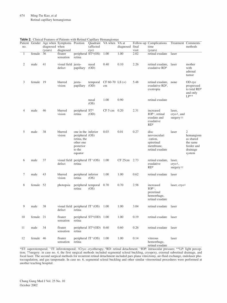
Table 2. Clinical Features of Patients with Retinal Capillary HemangiomasPatient Gender Age when Symptoms Position Quadrant VA when VA at Follow-up Complications Treatment Comments
no. diagnosed when (affected diagnosed final time methods(years) diagnosed eye) visit (years)
1 female 36 floater peripheral ST*(OS) 1.00 1.00 2.02 retinal exudate lasersensation retina
2 male 41 visual field juxta- nasal 0.40 0.10 2.26 retinal exudate, laser mother defect papillary (OD) exudative RDII with
adrenaltumor
3 female 19 blurred juxta- temporal CF 60-70 LS (+) 5.48 retinal exudate, none OD eye vision papillary (OD) cm exudative RDII, progressed
exotropia to total RDII
and only LP**
nasal 1.00 0.90 retinal exudate(OS)
4 male 46 blurred peripheral ST* CF 5 cm 0.20 2.31 increased laser, vision retina (OD) IOP£k, retinal cryo , and
exudate and surgeryexudativeRDII
5 male 38 blurred one in the inferior 0.03 0.01 0.27 disc laser 2 vision peripheral (OS) neovasculari hemangiom
retina, the -zation, as sharedother one epiretinal the same posterior membrane, feeder andto the retinal exudate drainageequator system
6 male 37 visual field peripheral IT (OS) 1.00 CF 25cm 2.73 retinal exudate, laser,defect retina exudative cryo ,
RDII surgery
7 male 43 blurred peripheral inferior 1.00 1.00 0.62 retinal exudate laservision retina (OS)
8 female 52 photopsia peripheral temporal 0.70 0.70 2.58 increased laser, cryoretina (OS) IOP£k,
preretinalhemorrhage,retinal exudate
9 male 38 visual field peripheral IT (OS) 1.00 1.00 3.04 retinal exudate laserdefect retina
10 female 21 floater peripheral ST*(OD) 1.00 1.00 0.19 retinal exudate lasersensation retina
11 male 34 floater peripheral ST*(OD) 0.60 0.60 0.26 retinal exudate lasersensation retina
12 female 46 floater peripheral IT (OS) 1.00 1.00 0.14 vitreous lasersensation retina hemorrhage,
retinal exudate*ST: superotemporal; IT: inferotemporal; Cryo: cryotherapy; IIRD: retinal detachment; £kIOP: intraocular pressure; **LP: light percep-tion; ??surgery: in case no. 4, the first surgical methods included segmental scleral buckling, cryopexy, external subretinal drainage, andfocal laser. The second surgical methods for recurrent retinal detachment included pars plana vitrectomy, air-fluid exchange, endolaser pho-tocoagulation, and gas tamponade. In case no. 6, segmental scleral buckling and other similar vitreoretinal procedures were performed atanother teaching hospital.

Chang Gung Med J Vol. 25 No. 10October 2002
Ming-Tse Kuo, et alRetinal capillary hemangiomas
675
cryotherapy. One patient (no. 4) received segmentalscleral buckling, cryotherapy, external subretinaldrainage, and focal laser at the first operation. Dueto recurrent retinal detachment, a second operationconsisting of pars plana vitrectomy, air-fluidexchange, endolaser photocoagulation, and gas tam-ponade was performed after 1 month. The otherpatient (no. 6) received laser treatment and cryother-apy, but these failed in our hospital, and that patientreceived segmental scleral buckling and other similarvitreoretinal procedures at another teaching hospital.Two patients with juxtapapillary hemangiomas hadexudative retinal detachment at their first visit. Onepatient (patient no. 3) received close observationwithout laser or any other treatment, and her visualacuity at the final visit was light perception only.The other patient (no. 2) received repeated focallaser, and his visual acuity at the final visit was 0.1.In the 12 patients, the major ocular complicationsincluded retinal exudation and exudative retinaldetachment. All patients suffered from retinal exu-dation, and 4 patients suffered from exudative retinaldetachment. Other ocular complications included 2patients with increased intraocular pressure, 1 patient
with an epiretinal membrane, 1 patient with prereti-nal hemorrhage, 1 patient with vitreous hemorrhage,1 patient with neovascularization of the disc, and 1patient with exotropia (Table 2). The pedigree focus-ing on ocular problems, central nervous systemtumors, renal tumors, and other visceral lesions wastaken by chart review and was followed up with tele-phone interviews if the records were incomplete.Seven patients received computed tomography of thebrain, and 2 patients received renal ultrasound scans.According to the pedigree and systemic surveys, nopatient met the diagnostic criteria of von Hippel-Lindau disease.
Representative cases from the study aredescribed below.
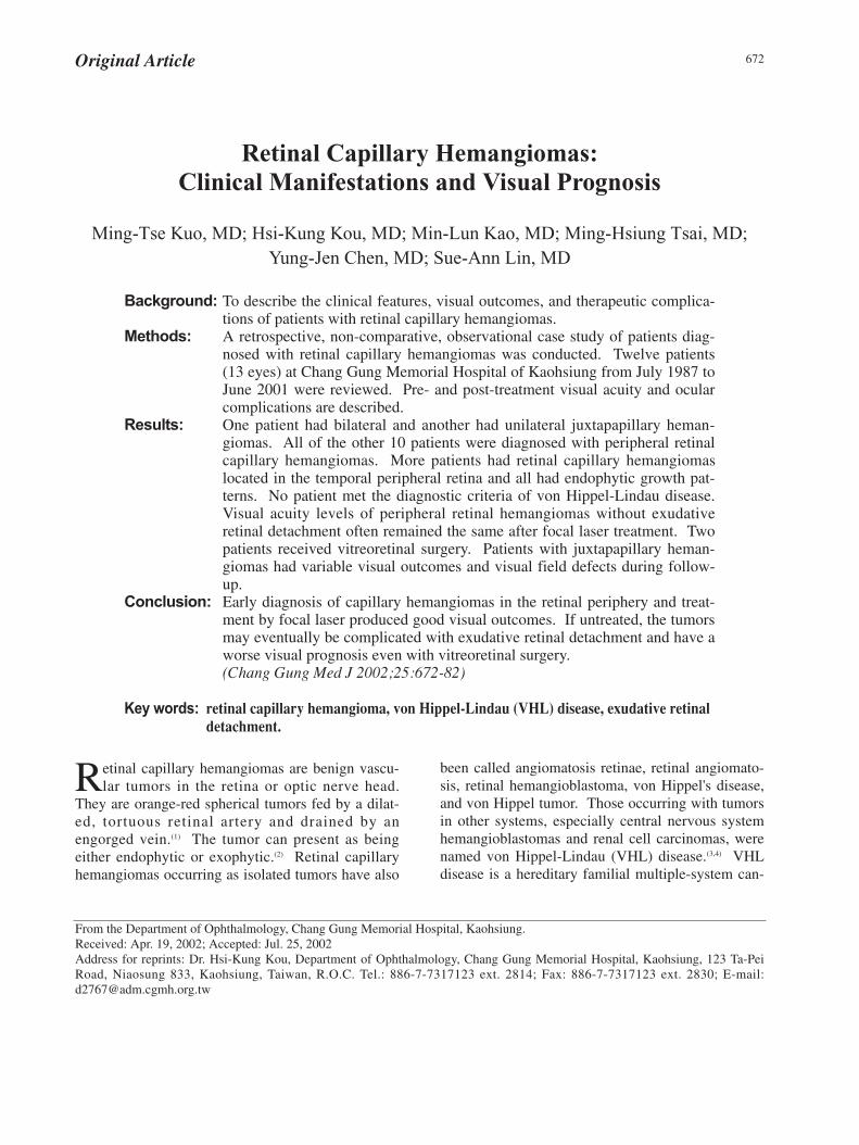
Patient no. 10A 21-year-old woman complained of a floater
sensation in the right eye for about 2 months. Initialvisual acuity was 1.0 in the right eye. Dilated fundusexamination revealed an endophytic tumor with adilated tortuous artery and vein in the superotempo-ral peripheral retina. Retinal exudates surroundedthe tumor (Fig. 2A). Fluorescein angiographyshowed an early filling of the fine tumor capillaries,hyperfluorescence of the retinal hemangioma withdilated feeder arteries and drainage veins, andintense late staining (Fig. 2B). After repeated focallaser treatment, the tumor shrank, and the diameterof the feeder arteries and drainage veins decreased(Fig. 2C). Her visual acuity remained at 1.0 at thefinal visit.
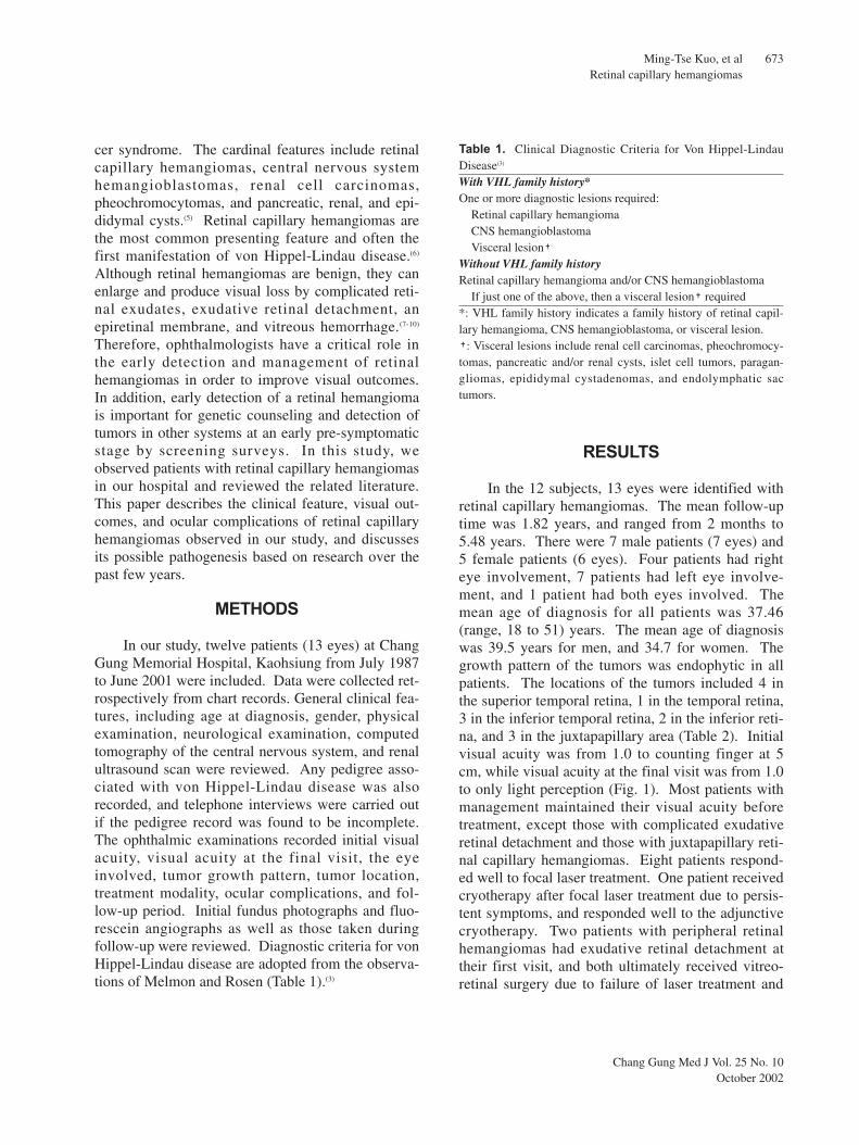
Patient no. 6A 37-year-old man suffered from superior visual
field defect in the left eye for about 3 weeks. Initialvisual acuity was 1.0 in the left eye. Dilated fundusexamination showed severe lipid exudation over theinferior retina, and an endophytic red tumor withlocalized exudative retinal detachment in the infer-otemporal retina (Fig. 3A). Fluorescein angiographyconfirmed the diagnosis of retinal capillary heman-gioma (Fig. 3B). After repeated laser treatment, reti-nal exudation and exudative retinal detachment werestill progressing. Therefore, the patient received vit-reoretinal surgery at another teaching hospital.Ultimately, the retina of the patient became dry, andhis visual acuity was counting fingers at about 25 cmat the final visit.
Fig. 1 Visual acuity at diagnosis compared with visual acuityat the final visit. The transverse axis represents the visual acu-ity at onset, and the longitudinal axis represents the visualacuity at the final visit after management. The number inparentheses (6) represents the number of patients at the samepoint, i.e., there are 6 patients who maintained a visual acuityof 1.0 after treatment. Most of the patients retained good visu-al acuity after management.
Chang Gung Med J Vol. 25 No. 10October 2002
Ming-Tse Kuo, et alRetinal capillary hemangiomas
676
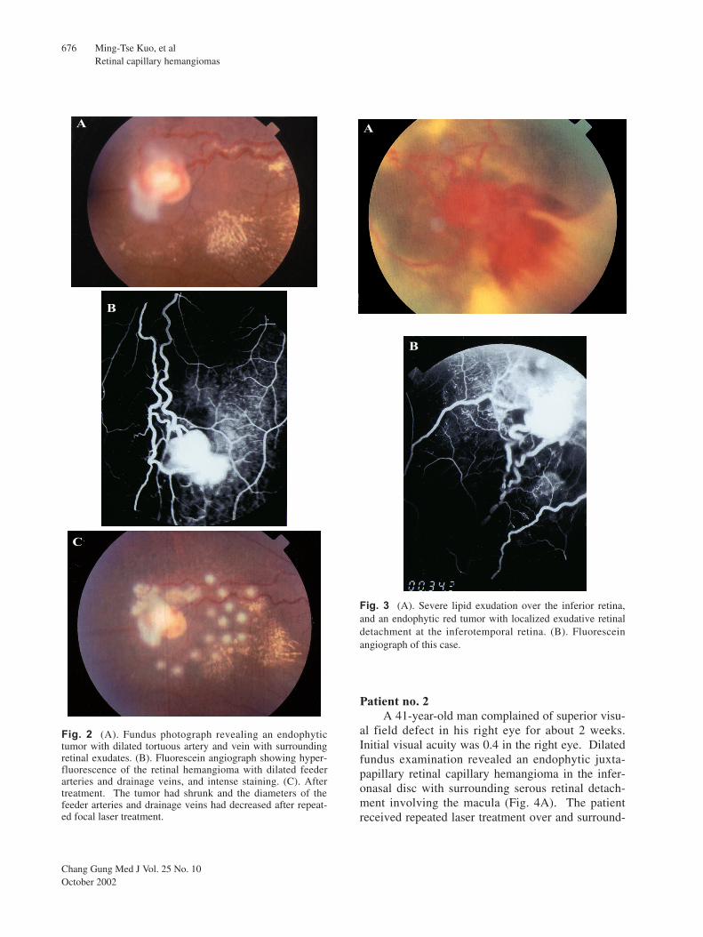
Patient no. 2A 41-year-old man complained of superior visu-
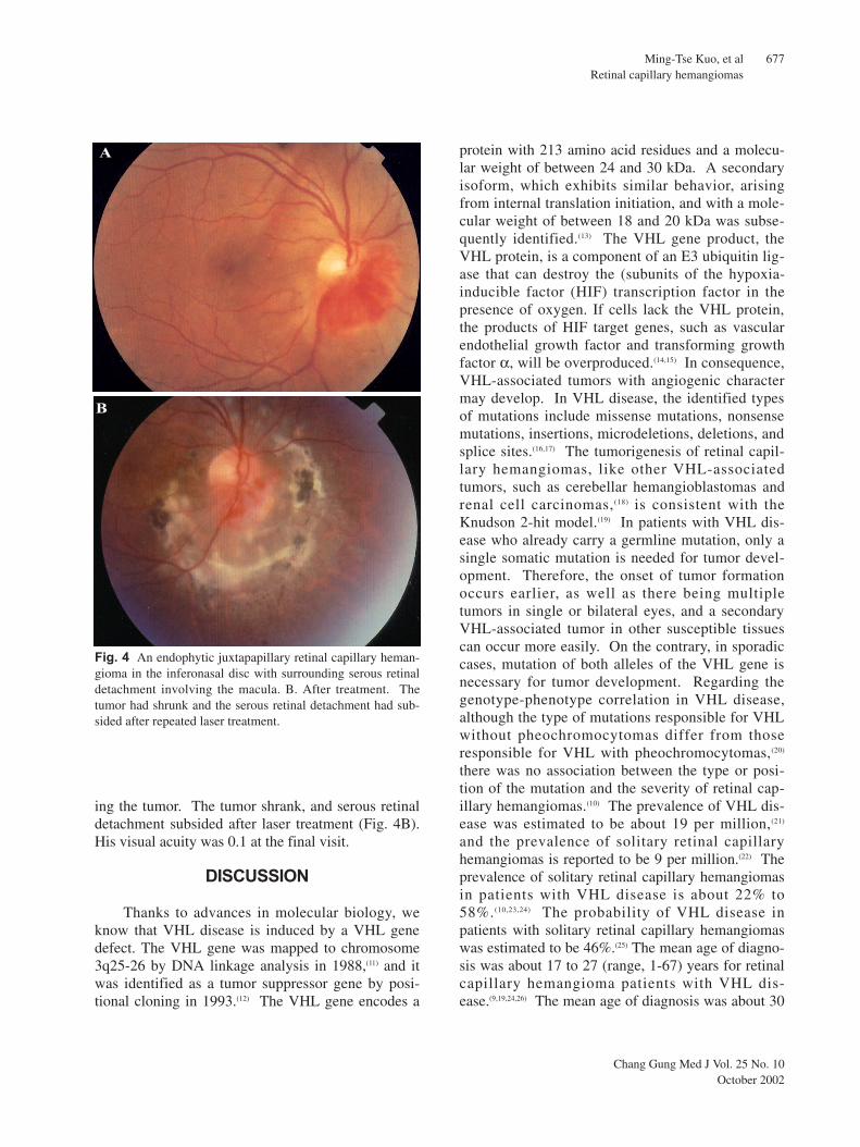
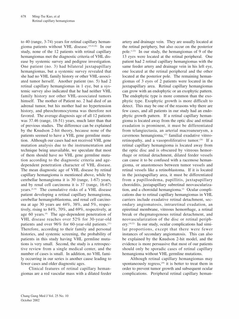
al field defect in his right eye for about 2 weeks.Initial visual acuity was 0.4 in the right eye. Dilatedfundus examination revealed an endophytic juxta-papillary retinal capillary hemangioma in the infer-onasal disc with surrounding serous retinal detach-ment involving the macula (Fig. 4A). The patientreceived repeated laser treatment over and surround-
Fig. 3 (A). Severe lipid exudation over the inferior retina,and an endophytic red tumor with localized exudative retinaldetachment at the inferotemporal retina. (B). Fluoresceinangiograph of this case.
B
A
B
C
Fig. 2 (A). Fundus photograph revealing an endophytictumor with dilated tortuous artery and vein with surroundingretinal exudates. (B). Fluorescein angiograph showing hyper-fluorescence of the retinal hemangioma with dilated feederarteries and drainage veins, and intense staining. (C). Aftertreatment. The tumor had shrunk and the diameters of thefeeder arteries and drainage veins had decreased after repeat-ed focal laser treatment.
A
Chang Gung Med J Vol. 25 No. 10October 2002
Ming-Tse Kuo, et alRetinal capillary hemangiomas
677
ing the tumor. The tumor shrank, and serous retinaldetachment subsided after laser treatment (Fig. 4B).His visual acuity was 0.1 at the final visit.
DISCUSSION
Thanks to advances in molecular biology, weknow that VHL disease is induced by a VHL genedefect. The VHL gene was mapped to chromosome3q25-26 by DNA linkage analysis in 1988,(11) and itwas identified as a tumor suppressor gene by posi-tional cloning in 1993.(12) The VHL gene encodes a
protein with 213 amino acid residues and a molecu-lar weight of between 24 and 30 kDa. A secondaryisoform, which exhibits similar behavior, arisingfrom internal translation initiation, and with a mole-cular weight of between 18 and 20 kDa was subse-quently identified.(13) The VHL gene product, theVHL protein, is a component of an E3 ubiquitin lig-ase that can destroy the (subunits of the hypoxia-inducible factor (HIF) transcription factor in thepresence of oxygen. If cells lack the VHL protein,the products of HIF target genes, such as vascularendothelial growth factor and transforming growthfactor α, will be overproduced.(14,15) In consequence,VHL-associated tumors with angiogenic charactermay develop. In VHL disease, the identified typesof mutations include missense mutations, nonsensemutations, insertions, microdeletions, deletions, andsplice sites.(16,17) The tumorigenesis of retinal capil-lary hemangiomas, like other VHL-associatedtumors, such as cerebellar hemangioblastomas andrenal cell carcinomas,(18) is consistent with theKnudson 2-hit model.(19) In patients with VHL dis-ease who already carry a germline mutation, only asingle somatic mutation is needed for tumor devel-opment. Therefore, the onset of tumor formationoccurs earlier, as well as there being multipletumors in single or bilateral eyes, and a secondaryVHL-associated tumor in other susceptible tissuescan occur more easily. On the contrary, in sporadiccases, mutation of both alleles of the VHL gene isnecessary for tumor development. Regarding thegenotype-phenotype correlation in VHL disease,although the type of mutations responsible for VHLwithout pheochromocytomas differ from thoseresponsible for VHL with pheochromocytomas,(20)
there was no association between the type or posi-tion of the mutation and the severity of retinal cap-illary hemangiomas.(10) The prevalence of VHL dis-ease was estimated to be about 19 per million,(21)
and the prevalence of solitary retinal capillaryhemangiomas is reported to be 9 per million.(22) Theprevalence of solitary retinal capillary hemangiomasin patients with VHL disease is about 22% to58%.(10,23,24) The probability of VHL disease inpatients with solitary retinal capillary hemangiomaswas estimated to be 46%.(25) The mean age of diagno-sis was about 17 to 27 (range, 1-67) years for retinalcapillary hemangioma patients with VHL dis-ease.(9,19,24,26) The mean age of diagnosis was about 30
A
B
Fig. 4 An endophytic juxtapapillary retinal capillary heman-gioma in the inferonasal disc with surrounding serous retinaldetachment involving the macula. B. After treatment. Thetumor had shrunk and the serous retinal detachment had sub-sided after repeated laser treatment.
Chang Gung Med J Vol. 25 No. 10October 2002
Ming-Tse Kuo, et alRetinal capillary hemangiomas
678
to 40 (range, 3-74) years for retinal capillary heman-gioma patients without VHL disease.(19,24,26) In ourstudy, none of the 12 patients with retinal capillaryhemangiomas met the diagnostic criteria of VHL dis-ease by systemic survey and pedigree investigation.One patient (no. 3) had bilateral juxtapapillaryhemangiomas, but a systemic survey revealed thatshe had no VHL family history or other VHL-associ-ated tumor herself. Another patient (no. 5) had 2retinal capillary hemangiomas in 1 eye, but a sys-temic survey also indicated that he had neither VHLfamily history nor other VHL-associated tumorshimself. The mother of Patient no. 2 had died of anadrenal tumor, but his mother had no hypertensionhistory, and pheochromocytoma was therefore notfavored. The average diagnosis age of all 12 patientswas 37.46 (range, 18-51) years, much later than thatof previous studies. The difference can be explainedby the Knudson 2-hit theory, because none of thepatients seemed to have a VHL gene germline muta-tion. Although our cases had not received VHL genemutation analysis due to the instrumentation andtechnique being unavailable, we speculate that mostof them should have no VHL gene germline muta-tion according to the diagnostic criteria and age-dependent penetration character of VHL disease.The mean diagnostic age of VHL disease by retinalcapillary hemangioma is mentioned above, while bycerebellar hemangioma it is 30 (range, 1-67) years,and by renal cell carcinoma it is 37 (range, 16-67)years.(6,18) The cumulative risks of a VHL diseasepatient developing a retinal capillary hemangioma,cerebellar hemangioblastoma, and renal cell carcino-ma at age 30 years are 44%, 38%, and 5%, respec-tively, rising to 84%, 70%, and 69%, respectively, atage 60 years.(9) The age-dependent penetration ofVHL disease reaches over 52% for 30-year-oldpatients and over 96% for 60-year-old patients.(21)
Therefore, according to their family and personalhistories, and systemic screening, the probability ofpatients in this study having VHL germline muta-tions is very small. Second, the study is a retrospec-tive review from a single medical center, and thenumber of cases is small. In addition, no VHL fami-ly occurring in our series is another cause leading tofewer cases and older diagnostic ages.
Clinical features of retinal capillary heman-giomas are a red vascular mass with a dilated feeder
artery and drainage vein. They are usually located atthe retinal periphery, but also occur on the posteriorpole.(1,22) In our study, the hemangiomas of 9 of the13 eyes were located at the retinal peripheral. Onepatient had 2 retinal capillary hemangiomas with thesame feeder artery and drainage vein in his left eye,one located at the retinal peripheral and the otherlocated at the posterior pole. The remaining heman-giomas of 3 eyes of 2 patients were located in thejuxtapapillary area. Retinal capillary hemangiomascan grow with an endophytic or an exophytic pattern.The endophytic type is more common than the exo-phytic type. Exophytic growth is more difficult todetect. This may be one of the reasons why there arefew cases, and all patients in our study had an endo-phytic growth pattern. If a retinal capillary heman-gioma is located away from the optic disc and retinalexudation is prominent, it must be differentiatedfrom telangiectasia, an arterial macroaneurysm, acavernous hemangioma,(27) familial exudative vitreo-retinopathy, and a vasoproliferative tumor.(28) If aretinal capillary hemangioma is located away fromthe optic disc and is obscured by vitreous hemor-rhage or retinal detachment, dilated feeder vesselscan cause it to be confused with a racemose heman-gioma, or anastomoses between tumor vessels andretinal vessels like a retinoblastoma. If it is locatedin the juxtapapillary area, it must be differentiatedfrom a papilloedema, papillitis, juxtapapillarychoroiditis, juxtapapillary subretinal neovasculariza-tion, and a choroidal hemangioma.(2) Ocular compli-cations due to retinal capillary hemangiomas in VHLcarriers include exudative retinal detachment, sec-ondary angiomatosis, intraretinal exudation, anepiretinal membrane, vitreous hemorrhage, a retinalbreak or rhegmatogenous retinal detachment, andneovascularization of the disc or retinal periph-ery.(10,22) In our study, ocular complications had simi-lar proportions, except that there were fewerinstances of secondary angiomatosis. This can alsobe explained by the Knudson 2-hit model, and theevidence is more persuasive that most of our patientsshould only be sporadic cases of retinal capillaryhemangioma without VHL germline mutations.
Although retinal capillary hemangiomas mayspontaneously regress,(29) it is better to treat them inorder to prevent tumor growth and subsequent ocularcomplications. Peripheral retinal capillary heman-
Chang Gung Med J Vol. 25 No. 10October 2002
Ming-Tse Kuo, et alRetinal capillary hemangiomas
679
giomas respond well to treatment especially if theyare small, but the outcome of treatment is still disap-pointing for juxtapapillary capillary hemangiomas.The mainstay of treatment is laser therapy. The kryp-ton laser or argon laser,(7,30) which can be wellabsorbed by hemoglobin, is a good choice. Largerlesions (greater than 3 mm in diameter), lesions inthe extreme periphery, and those with underlyingsubretinal fluid are well managed with cryothera-py.(7,8,31) Patients with juxtapapillary capillary heman-giomas treated by either observation or laser general-ly have variable visual acuity.(2,30,32) The treatment ofjuxtapapillary capillary hemangiomas by diodetranspupillary thermophotocoagulation remains con-troversial.(33,34) For patients with ocular complica-tions, such as retinal exudates, exudative retinaldetachment, epiretinal membrane formation, and vit-reous hemorrhage, vitreoretinal surgery is ultimatelyrequired, and blindness is common.(10,22,35) In ourstudy, for those patients with retinal capillary heman-gioma away from the disc, initial visual acuity was1.0 in 6 of 10 eyes, ≥ 0.4 in 8 of 10 eyes, and ≥ 0.1 in8 of 10 eyes. The visual acuity at the final visit was
1.0 in 5 of 10 eyes, ≥ 0.4 in 7 of 10 eyes, and ≥ 0.1 in8 of 10 eyes. Most of the patients retained goodvisual acuity after treatment, except for 2 patientswith a severe complicated exudative retinal detach-ment who ultimately received vitreoretinal surgery.In one of the patients with a juxtapapillary capillaryhemangioma who received laser treatment, initialvisual acuity was 0.4, while it was 0.1 at the finalfollow-up. For the other patient with bilateral juxta-papillary capillary hemangiomas who receivedobservation only, initial visual acuity was countingfinger at 60-70 cm for her right eye, and 1.0 for herleft eye. Visual acuity at the final visit was light per-ception only for her right eye, and 0.9 for her lefteye. The visual outcomes in our study are compara-ble with those of previous studies.(7,22)
Although none of our patients with retinal capil-lary hemangiomas were associated with VHL dis-ease, we should still pay more attention to the highcorrelation between these entities due to the highmorbidity and mortality of VHL disease.Ophthalmologists usually are the earliest detectors ofVHL disease. Therefore, once a retinal capillary
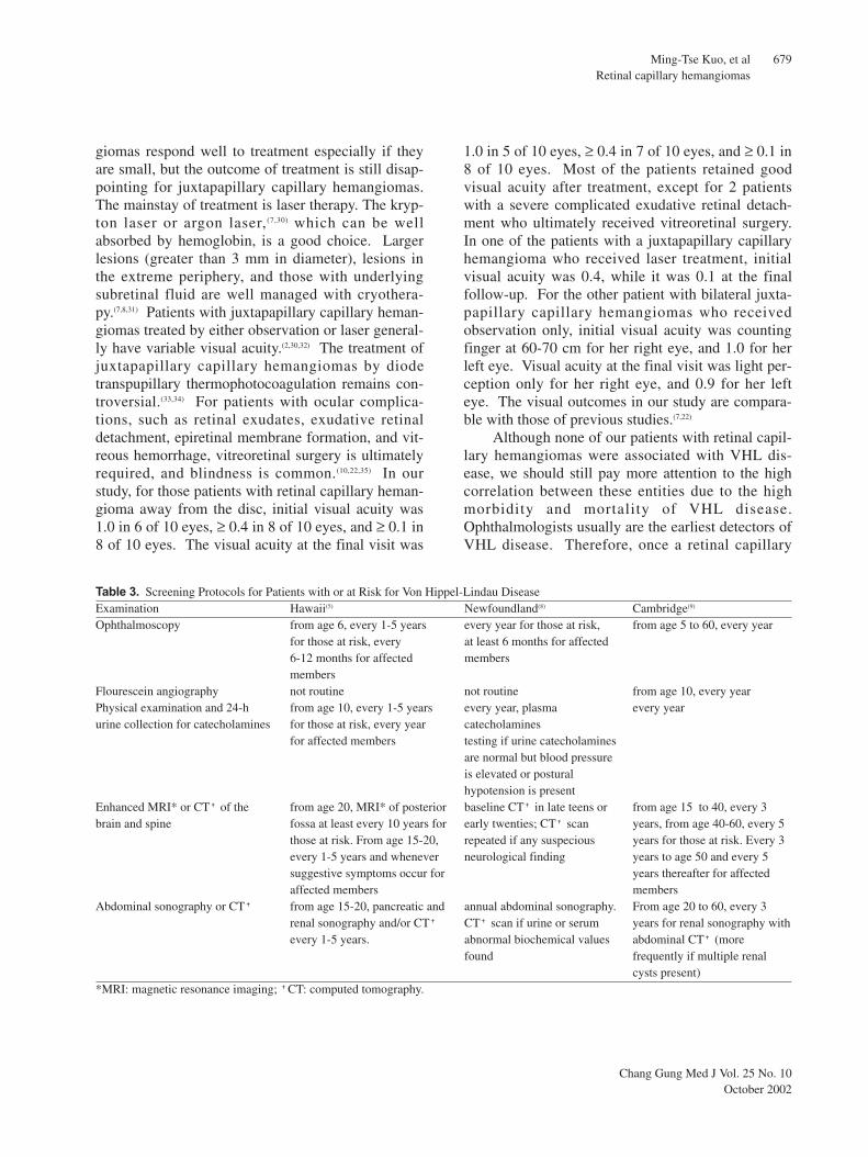
Table 3. Screening Protocols for Patients with or at Risk for Von Hippel-Lindau DiseaseExamination Hawaii(5) Newfoundland(8) Cambridge(9)
Ophthalmoscopy from age 6, every 1-5 years every year for those at risk, from age 5 to 60, every yearfor those at risk, every at least 6 months for affected 6-12 months for affected members members
Flourescein angiography not routine not routine from age 10, every yearPhysical examination and 24-h from age 10, every 1-5 years every year, plasma every yearurine collection for catecholamines for those at risk, every year catecholamines
for affected members testing if urine catecholaminesare normal but blood pressure is elevated or postural hypotension is present
Enhanced MRI* or CT of the from age 20, MRI* of posterior baseline CT in late teens or from age 15 to 40, every 3brain and spine fossa at least every 10 years for early twenties; CT scan years, from age 40-60, every 5
those at risk. From age 15-20, repeated if any suspecious years for those at risk. Every 3every 1-5 years and whenever neurological finding years to age 50 and every 5suggestive symptoms occur for years thereafter for affected affected members members
Abdominal sonography or CT from age 15-20, pancreatic and annual abdominal sonography. From age 20 to 60, every 3 renal sonography and/or CT CT scan if urine or serum years for renal sonography with every 1-5 years. abnormal biochemical values abdominal CT (more
found frequently if multiple renal cysts present)
*MRI: magnetic resonance imaging; CT: computed tomography.
Chang Gung Med J Vol. 25 No. 10October 2002
Ming-Tse Kuo, et alRetinal capillary hemangiomas
680
hemangioma is identified, a detailed family historyshould be obtained, and a subsequent systemicscreening protocol should be arranged to exclude afatal central nervous system (CNS) hemangioblas-toma and visceral lesions of VHL disease. There aredifferent screening protocols for VHL disease, butthey have similar items of examination includingophthalmoscopy, fluorescein angiography, brain andspine magnetic resonance imaging (MRI) or comput-ed tomography (CT), abdominal ultrasound or CT,and urinary catecholamine testing (Table 3).(5,8,9)
Screening regimens can be used for patients withknown VHL disease or for patients at risk for thisdisease to decrease their morbidity and mortality bypresymptomatic diagnosis and management. Genetictesting methods that combine quantitative Southernblot analysis for detecting deletions of the entireVHL gene, Southern blotting for detecting generearrangement, fluorescence in situ hybridization forconforming deletions, and complete sequencing ofthe gene have all contributed to high detectionrates.(36) If molecular biological techniques andequipment are available, DNA testing can be per-formed to exclude the diagnosis of VHL disease withsolitary retinal capillary hemangiomas, to assess therisk of specific mutations in patients with knownVHL disease, to screen at-risk relatives, and to doprenatal screening after detailed genetic counsel-ing.(37,38) None of the 12 patients in the study met theclinical diagnostic criteria of VHL disease, and wehope that the availability of genetic testing in thefuture can further confirm the diagnosis without thegermline mutation of the VHL gene.
In summary, early diagnosis of capillary heman-giomas in the retinal periphery and treatment byfocal laser produced good visual outcomes.Cryotherapy is an alternative or adjunctive manage-ment for specific situations like larger tumors orthose on the extreme peripheral retina. Without treat-ment or with delayed treatment, capillary heman-giomas in the retinal periphery may be complicatedwith severe exudation and exudative retinal detach-ment, and produce a worse visual prognosis evenwith vitreoretinal surgery. For patients with juxta-papillary retinal capillary hemangiomas, laser treat-ment should be more prudent due to the variablevisual outcomes. Transpupillary thermophotocoagu-lation is still controversial for the treatment of juxta-
papillary retinal capillary hemangiomas. Better man-agement of juxtapapillary hemangiomas is mandato-ry, and needs to be determined with further study.
REFERENCES
1. Goldberg MF, Duke JR. Histopathologic findings in atreated and an untreated eye. Am J Ophthalmol 1968;66:693-705.
2. Gass JDW, Braunstein R. Sessile and exophytic capillaryangiomas of the juxtapapillary retina and optic nervehead. Arch Ophthalmol 1980;98:1790-7.
3. Melmon KL, Rosen SW. Lindau's disease. Am J Med1964;36:595-617.
4. Horton WA, Wong V, Eldridge R. Von Hippel Lindau dis-ease. Clinical and pathological manifestations in ninefamilies with 50 affected members. Arch Int Med1976;136:769-77.
5. Lamiell JM, Salazar FG, Hsia YE. Von Hippel-Lindaudisease affecting 43 members of a single kindred.Medicine (Baltimore) 1989;68:1-29.
6. Choyke PL, Glenn GM, McClellan M. Von Hippel-Lindau disease: genetic, clinical and imaging feature.Radiology 1995;146:629-42.
7. Annesley WH, Leonard BC, Shields JA, Tasman WS.Fifteen year review of treated cases of retinal angiomato-sis. Trans Am Acad Ophthalmol Otolaryngol 1977;83:446-53.
8. Ridley M, Green J, Johnson G. Retinal angiomatosis: theocular manifestations of von Hippel-Lindau disease. CanJ Ophthalmol 1986;21:276-83.
9. Maher ER, Yates JR, Harries R, Benjamin C, Harris R,Moore AT. Clinical features & nature history of vonHippel-Lindau disease. Quart J Med 1990;77:1151-63.
10. Webster A, Maher E, Moore A. Clinical characteristics ofocular angiomatosis in von Hippel-Lindau disease andcorrelation with germline mutation. Arch Ophthalmol1999;117:371-8.
11. Seizinger BR, Rouleau GA, Ozelius LJ, Lane AH, FarmerGE, Lamiell JM. Von Hippel-Lindau disease maps to theregion of chromosome 3 associated with renal cell carci-noma. Nature 1988;332:268-9.
12. Latif F, Tory K, Gnarra J. Identification of the VonHippel-Lindau dz tumor suppressor gene. Science1993;260:1317-20.
13. Iliopoulos O, Ohh M, Kaelin W. pVHL 19 is a biological-ly active product of the von Hippel-Lindau gene arisingfrom internal translation initiation. Proc Natl Acad SciUSA 1998;95:11661-6.
14. Kondo K, Kaelin WG. The von Hippel-Lindau tumor sup-pressor gene. Exp Cell Res 2001:264;117-25.
15. de Paulsen N, Brychzy A, Fournier MC, Klausner RD,Gnarra JR, Pause A, Lee S. Role of transforming growthfactor-alpha in von Hippel-Lindau (VHL)(-/-) clear cell
Chang Gung Med J Vol. 25 No. 10October 2002
Ming-Tse Kuo, et alRetinal capillary hemangiomas
681
renal carcinoma cell proliferation: a possible mechanismcoupling VHL tumor suppressor inactivation and tumori-genesis. Proc Natl Acad Sci USA 2001;98:1387-92.
16. Richards FM, Crossey PA, Phipps ME. Detailed mappingof germline deletions of the von Hippel-Lindau diseasetumour suppressor gene. Hum Mol Genet 1994; 3: 595-8.
17. Zbar B, Kishida T, Chen F, Schmidt L, Maher ER,Richards FM, Crossey PA, Webster AR, Affara NA,Ferguson Smith MA, Brauch H, Glavac D, Neumann HP,Tisherman S, Mulvihill JJ, Gross DJ, Shuin T, Whaley J,Seizinger B, Kley N, Olschwang S, Boisson C, Richard S,Lips CH, Lerman M. Germline mutations in the vonHippel-Lindau disease (VHL) gene in families fromNorth America, Europe, and Japan. Hum Mutat 1996;8:348-57.
18. Maher ER, Yates JR, Ferguson-Smith MA. Statisticalanalysis of the two-stage mutation model in von Hippel-Lindau disease, and in sporadic cerebellar haemangioblas-toma and renal cell carcinoma. J Med Genet 1990;27:311-4.
19. Chang JH, Spraul CW, Lynn ML, Drack A, GrossniklausHE. The two-stage mutation model in retinal heman-gioblastoma. Ophthalmic Genet 1998;19:123-30.
20. Chen F, Kishida T, Yao M. Germ line mutations in thevon Hippel-Lindau disease tumor suppressor gene: corre-lations with phenotype. Hum Mutat 1995;5:66-75.
21. Maher ER, Iselius L, Yates JRW, Littler M, Benjamin C,Harris R. Von Hippel-Lindau disease: a genetic study. JMed Genet 1991;28:443-7.
22. Webster A, Maher E, Bird A, Gregor Z, Moore. A clinicaland molecular genetic analysis of solitary ocular angioma.Ophthalmology 1999;106:623-9.
23. Kreusel KM, Bechrakis NE, Heinichen T, Neumann L,Neumann HP, Foerster MH. Retinal angiomatosis and vonHippel-Lindau disease. Graefe's Arch Clin ExpOphthalmol 2000;238:916-21.
24. Niemela M, Lemeta S, Sainio M, Rauma S, Pukkala E,Kere J, Bohling T, Laatikainen L, Jaaskelainen J,Summanen P. Hemangioblastomas of the retina: impact ofvon Hippel-Lindau disease. Invest Ophthalmol Vis Sci:2000;41:1909-15.
25. Singh A, Shields J, Shields C. Solitary retinal capillaryhemangioma: hereditary (von Hippel-Lindau disease) ornonhereditary ? Arch Ophthalmol 2001;119:232-4.
26. Singh AD, Nouri M, Shields CL, Shields JA, Smith AF.
Retinal capillary hemangioma. A comparison of sporadiccases and cases associated with von Hippel-Lindau dis-ease. Ophthalmology 2001;108:1907-11.
27. Gass JDM. Cavernous haemangioma of the retina. Am JOphthalmol 1971;71:799-814.
28. Shields CL, Shields JA, Barrett J, De Potter P.Vasoproliferative tumors of the ocular fundus.Classification and clinical manifestations in 103 patients.Arch Ophthalmol 1995;113:615-23.
29. Whitson DJ, Welch RB. Von Hippel-Lindau disease: casereport of a patient with spontaneous regression of a retinalangioma. Retina 1986;6:253-9.
30. Kremer I, Gilad E, Ben-Sira I. Juxtapapillary exophyticcapillary hemangioma treated by yellow krypton (568nm) laser photocoagulation. Ophthalmic Surg1988:19:743-7.
31. Shields JA. Response of retinal capillary hemangioma tocryotherapy. Arch Ophthalmology 1993;111:551.
32. McCabe CM, Flynn HW Jr, Shields CL, Shields JA,Regillo CD, McDonald HR, Berrocal MH, Gass JD,Mieler WF. Juxtapapillary capillary hemangiomas.Clinical features and visual acuity outcomes.Ophthalmology 2000;107:2240-8.
33. Transpupillary thermotherapy for retinal capillary heman-gioma in von Hippel-Lindau disease. Parmar DN,Mireskandari9 K, McHugh D. Ophthalmic Surgery &Lasers 2000;31:334-6.
34. Garcia-Arumi J, Sararols LH, Cavero L, Escalada F,Corcostegui BF. Therapeutic options for capillary papil-lary hemangiomas. Ophthalmology 2000;107:48-54.
35. McDonald HR, Schatz H, Johnson RN, Abrams GW,Brown GC, Brucker AJ, Han DP, Lewis H, Mieler WF,Meyers S. Vitrectomy in eyes with peripheral retinalangioma associated with traction macular detachment.Ophthalmology 1996;103:329-35.
36. Stolle C, Glenn G, Zbar B. Improved detection ofgermline mutations in von Hippel-Lindau disease tumorsuppressor gene. Hum Mutat 1998;12:417-23
37. Geller G, Botkin JR, Green MJ. Genetic testing for sus-ceptibility to adult-onset cancer: the process and contentof informed consent. JAMA 1997;277:1467-74.
38. Levy M, Richard S. Attitudes of von Hippel-Lindau dis-ease patients towards presymptomatic genetic diagnosis inchildren prenatal diagnosis. J Med Genet 2000;37:476-8.
1987 2001 12(13 )
1210
1212 von Hippel-Lindau disease
2
( 2002;25:672-82)
von Hippel-Lindau (VHL) disease
“ł'‹'´ | “¶fl|ˇ †·‹¤⁄⁄Ø·̀¡G¥̀ Œ 91ƒ~4⁄º19⁄Ø¡F¥̀ Œ 91ƒ~7⁄º25⁄Ø¡Cfl̀ ¤œ' ƒL¥»‡B¡G‡¢¿fi¥́ fiv¡A“ł'‹'´ | †·‹¡C“¶fl¿⁄‡“Q¶m⁄jæ‚ 123‚„¡C Tel.: (07)7317123´ 2814; Fax: (07)7317123´ 2830; E-mail: [email protected]
682