pedi hemangioma pic 2014 03
TRANSCRIPT
8/17/2019 Pedi Hemangioma Pic 2014 03
http://slidepdf.com/reader/full/pedi-hemangioma-pic-2014-03 1/92
Hemangiomas in the
Pediatric Head and Nec
Review and UpdateResident Physician: Eugene Son, MD
Faculty Advisor: Harold Pine, MD, FAAP, FACS
The University of Texas Medical Branch
Department of Otolaryngology
Grand Rounds Presentation
March 28, 2014
Series Editor: Francis B. Quinn, Jr., MD, FACS – Archivist: Melinda Stoner Q
8/17/2019 Pedi Hemangioma Pic 2014 03
http://slidepdf.com/reader/full/pedi-hemangioma-pic-2014-03 2/92
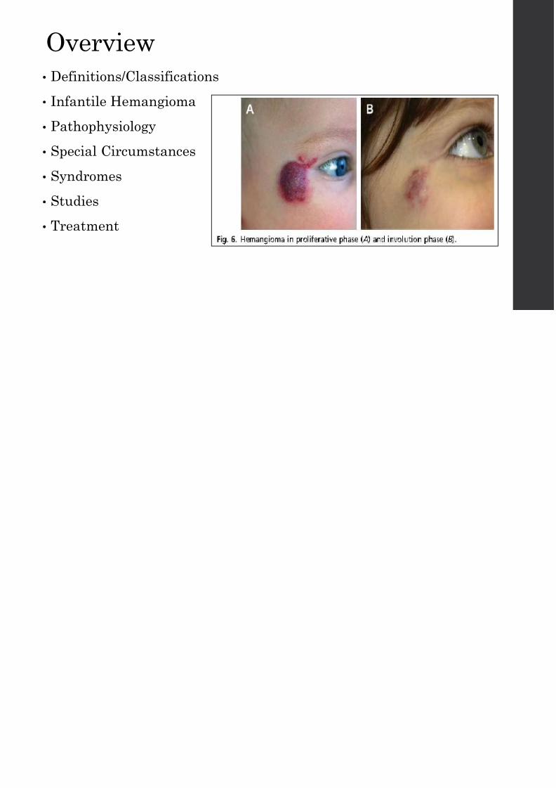
Overview
• Definitions/Classifications
• Infantile Hemangioma
• Pathophysiology
• Special Circumstances
• Syndromes
• Studies
• Treatment

8/17/2019 Pedi Hemangioma Pic 2014 03
http://slidepdf.com/reader/full/pedi-hemangioma-pic-2014-03 3/92
Definitions and
Classifications
8/17/2019 Pedi Hemangioma Pic 2014 03
http://slidepdf.com/reader/full/pedi-hemangioma-pic-2014-03 4/92
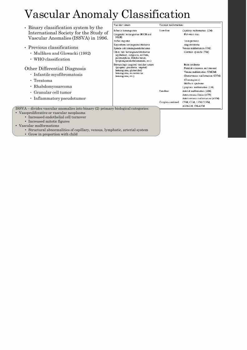
Vascular Anomaly Classification• Binary classification system by the
International Society for the Study of Vascular Anomalies (ISSVA) in 1996.
• Previous classifications
Mulliken and Glowacki (1982)
WHO classification
Other Differential Diagnosis
Infantile myofibromatosis
Teratoma
Rhabdomyosarcoma Granular cell tumor
Inflammatory pseudotumor
ISSVA – divides vascular anomalies into binary (2) primary biological categories:
• Vasoproliferative or vascular neoplasms
• Increased endothelial cell turnover
• Increased mitotic figures
• Vascular malformations
• Structural abnormalities of capillary, venous, lymphatic, arterial system• Grow in proportion with child
8/17/2019 Pedi Hemangioma Pic 2014 03
http://slidepdf.com/reader/full/pedi-hemangioma-pic-2014-03 5/92
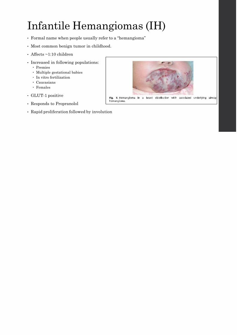
Infantile Hemangiomas (IH)• Formal name when people usually refer to a “hemangioma”
• Most common benign tumor in childhood.
• Affects ~1:10 children
• Increased in following populations:
Premies
Multiple gestational babies
In vitro fertilization
Caucasians
Females
• GLUT-1 positive
• Responds to Propranolol
• Rapid proliferation followed by involution
8/17/2019 Pedi Hemangioma Pic 2014 03
http://slidepdf.com/reader/full/pedi-hemangioma-pic-2014-03 6/92
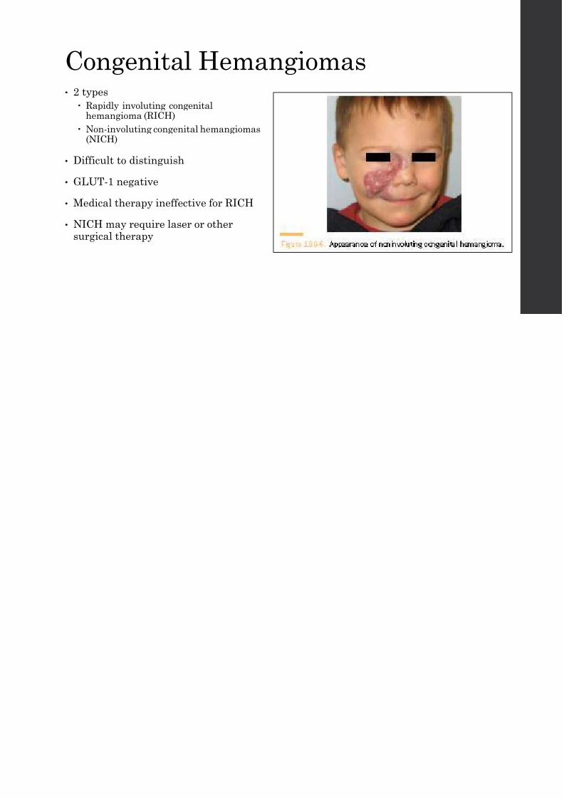
Congenital Hemangiomas• 2 types
Rapidly involuting congenitalhemangioma (RICH)
Non-involuting congenital hemangiomas(NICH)
• Difficult to distinguish
• GLUT-1 negative
• Medical therapy ineffective for RICH
• NICH may require laser or othersurgical therapy

8/17/2019 Pedi Hemangioma Pic 2014 03
http://slidepdf.com/reader/full/pedi-hemangioma-pic-2014-03 7/92
Lobular Capillary Hemangioma• AKA “ pyogenic granuloma”
• Etiology unknown, some evidence shows may be trauma or hormonalinfluences.
• Higher frequency on anterior nasal septum or anterior aspect of inferiorturbinates.
• More common in adults, especially pregnant females.
8/17/2019 Pedi Hemangioma Pic 2014 03
http://slidepdf.com/reader/full/pedi-hemangioma-pic-2014-03 8/92
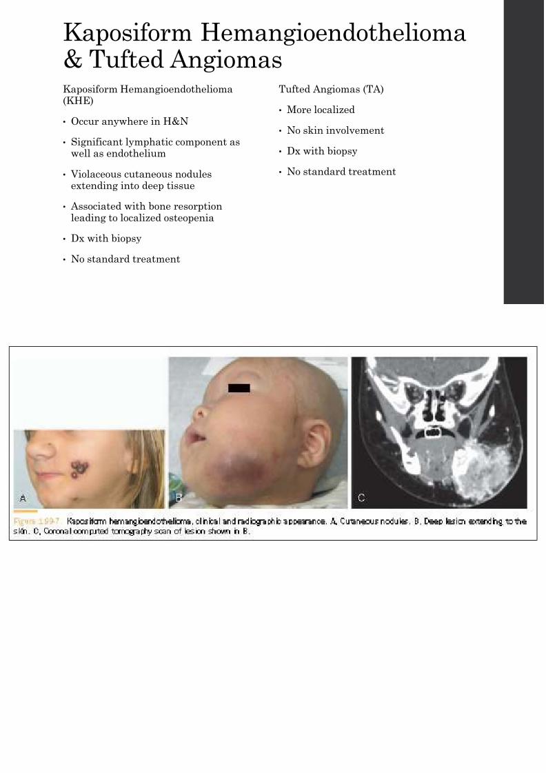
Kaposiform Hemangioendotheliom& Tufted AngiomasKaposiform Hemangioendothelioma(KHE)
• Occur anywhere in H&N
• Significant lymphatic component aswell as endothelium
• Violaceous cutaneous nodulesextending into deep tissue
• Associated with bone resorptionleading to localized osteopenia
• Dx with biopsy
• No standard treatment
Tufted Angiomas (TA)
• More localized
• No skin involvement
• Dx with biopsy
• No standard treatment

8/17/2019 Pedi Hemangioma Pic 2014 03
http://slidepdf.com/reader/full/pedi-hemangioma-pic-2014-03 9/92

8/17/2019 Pedi Hemangioma Pic 2014 03
http://slidepdf.com/reader/full/pedi-hemangioma-pic-2014-03 10/92
InfantileHemangioma (IH)
Overview
8/17/2019 Pedi Hemangioma Pic 2014 03
http://slidepdf.com/reader/full/pedi-hemangioma-pic-2014-03 11/92
Diagnosis•
Best diagnosed with thorough history and physicalexamination.
• Generally not present at birth
• Cutaneous abnormality in form of pallor, duskiness,telangiectasias.
• Bright red macule or papule rash with clear boundaries
• Color may deepen with time
• 5 or more cutaneous hemangiomas Recommend abdominal MRI (Liver)
8/17/2019 Pedi Hemangioma Pic 2014 03
http://slidepdf.com/reader/full/pedi-hemangioma-pic-2014-03 12/92
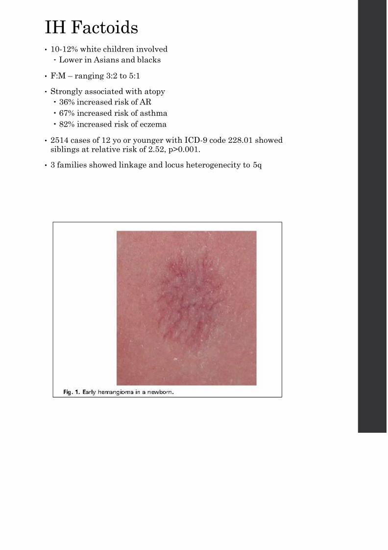
IH Factoids• 10-12% white children involved
Lower in Asians and blacks
• F:M – ranging 3:2 to 5:1
• Strongly associated with atopy
36% increased risk of AR
67% increased risk of asthma
82% increased risk of eczema
• 2514 cases of 12 yo or younger with ICD-9 code 228.01 showedsiblings at relative risk of 2.52, p>0.001.
• 3 families showed linkage and locus heterogenecity to 5q
8/17/2019 Pedi Hemangioma Pic 2014 03
http://slidepdf.com/reader/full/pedi-hemangioma-pic-2014-03 13/92
8/17/2019 Pedi Hemangioma Pic 2014 03
http://slidepdf.com/reader/full/pedi-hemangioma-pic-2014-03 14/92
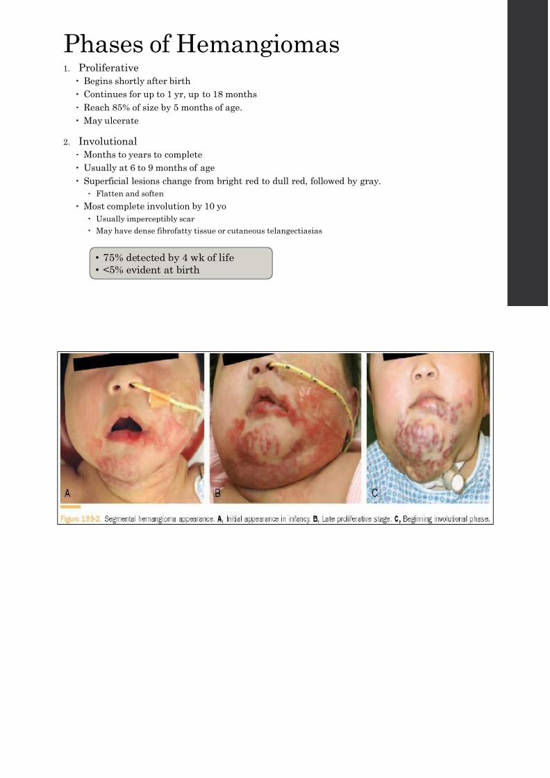
Phases of Hemangiomas1. Proliferative
Begins shortly after birth
Continues for up to 1 yr, up to 18 months Reach 85% of size by 5 months of age.
May ulcerate
2. Involutional
Months to years to complete
Usually at 6 to 9 months of age
Superficial lesions change from bright red to dull red, followed by gray.
Flatten and soften
Most complete involution by 10 yo
Usually imperceptibly scar
May have dense fibrofatty tissue or cutaneous telangectiasias
• 75% detected by 4 wk of life
• <5% evident at birth
8/17/2019 Pedi Hemangioma Pic 2014 03
http://slidepdf.com/reader/full/pedi-hemangioma-pic-2014-03 15/92
8/17/2019 Pedi Hemangioma Pic 2014 03
http://slidepdf.com/reader/full/pedi-hemangioma-pic-2014-03 16/92
Classification of IH
By depth of penetration
• Superficial lesions
Upper dermis
Strawberry red lesions
• Deep lesions
Dermal and subcutaneous tissue
Appear darker and more palpableas a mass
By location
• Focal
• Segmental
Larger and widespread

8/17/2019 Pedi Hemangioma Pic 2014 03
http://slidepdf.com/reader/full/pedi-hemangioma-pic-2014-03 17/92
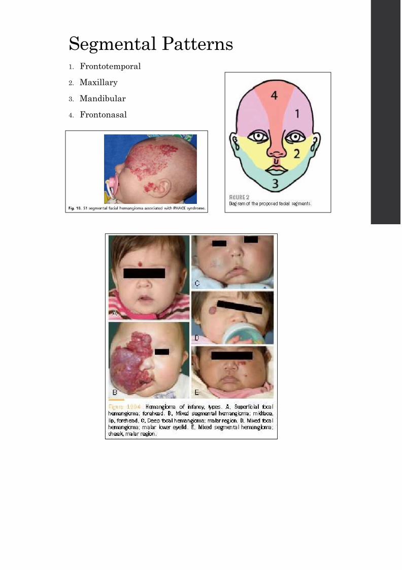
Segmental Patterns1. Frontotemporal
2. Maxillary
3. Mandibular
4. Frontonasal

8/17/2019 Pedi Hemangioma Pic 2014 03
http://slidepdf.com/reader/full/pedi-hemangioma-pic-2014-03 18/92
8/17/2019 Pedi Hemangioma Pic 2014 03
http://slidepdf.com/reader/full/pedi-hemangioma-pic-2014-03 19/92
Pathophysiology• Aberrant angiogenesis – formation of new blood vessels from preexisting
vasculature.
• Aberrant vasculogenesis – formation of new blood vessels from progenitorcells
• Dysregulated differentiation of embryonic cells. Isolated primitive mesenchymal cells CD133+ - human stem and progenitor cells
Form GLUT-1 positive vessels within 7-14 days
Cells differentiate into endothelium, adipocytes, pericytes.
• Molecular receptor pathways Vascular endothelial growth factor (VEGF)
B-adrenergic receptor pathway
Glucose transporter 1 (GLUT-1)
• Theories related to some hemangioma-placental connection
8/17/2019 Pedi Hemangioma Pic 2014 03
http://slidepdf.com/reader/full/pedi-hemangioma-pic-2014-03 20/92
Molecular Receptor Pathways• Vascular endothelial growth factor (VEGF)
Common mechanism for angiogenesis in tumors
Endothelial cells are derived from hemangioma stem cells Produce VEGF-A -> bind to VEGF receptors on these stem cells and stimulate
angiogenesis and differentiation into aberrant endothelial cells.
In vitro studies – hypoxia and estrogen synergistically enhance hemangiomaproliferation.
• Beta-adrenergic receptor pathway
Propranolol
Hypothesis
B-adrenergic vasoconstriction -> decreased bulk and flow
Ultrasound showed decrease in lesion volume and vessel density after therapyinitiation
Direct effect on apoptosis in capillary endothelial cells.
Down-regulates VEGF-A and HIF-1-alpha -> direct cytotoxic effect in the formof decreased endothelial cell migration and apoptosis.
8/17/2019 Pedi Hemangioma Pic 2014 03
http://slidepdf.com/reader/full/pedi-hemangioma-pic-2014-03 21/92
Molecular Receptor PathwaysOthers:
• Angiopoietins
ANG-1
ANG-2
• Tyrosine protein kinase receptors (Tie2)
• Notch Pathway
Notch 1-4 Delta-like 1, 3, 4
Jagged 1, 2
• Mammalian target of rapamycin complex (mTOR)

8/17/2019 Pedi Hemangioma Pic 2014 03
http://slidepdf.com/reader/full/pedi-hemangioma-pic-2014-03 22/92
GLUT-1• Glucose transporter 1 (GLUT-1)
• 97% of Hemangiomas test positive for GLUT-1
• Normally only in placental blood vessels and blood-brain barrier (BBB)blood vessels
• GLUT-1 negative Hemangiomas
• Noninvoluting congenital hemangiomas (NICH)
• Rapidly involuting congenital hemangiomas (RICH)
• Do not follow typical clinical course
8/17/2019 Pedi Hemangioma Pic 2014 03
http://slidepdf.com/reader/full/pedi-hemangioma-pic-2014-03 23/92
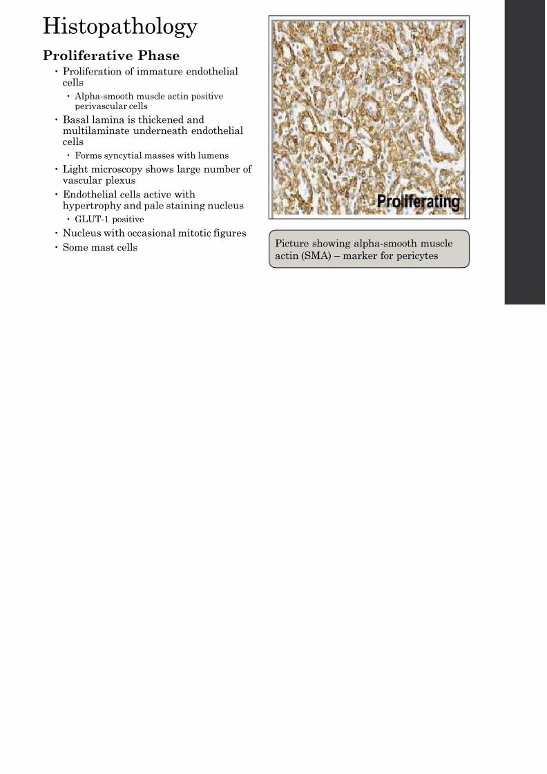
Histopathology
Proliferative Phase Proliferation of immature endothelial
cells
Alpha-smooth muscle actin positiveperivascular cells
Basal lamina is thickened andmultilaminate underneath endothelialcells
Forms syncytial masses with lumens
Light microscopy shows large number ofvascular plexus
Endothelial cells active withhypertrophy and pale staining nucleus
GLUT-1 positive
Nucleus with occasional mitotic figures
Some mast cells Picture showing alpha-smooth mu
actin (SMA) – marker for pericytes
8/17/2019 Pedi Hemangioma Pic 2014 03
http://slidepdf.com/reader/full/pedi-hemangioma-pic-2014-03 24/92
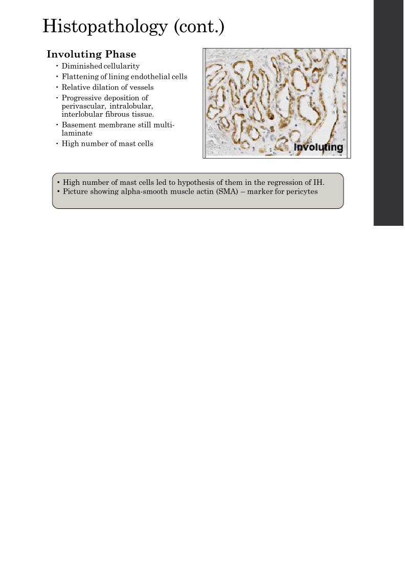
Histopathology (cont.)
Involuting Phase Diminished cellularity
Flattening of lining endothelial cells
Relative dilation of vessels
Progressive deposition ofperivascular, intralobular,interlobular fibrous tissue.
Basement membrane still multi-laminate
High number of mast cells
• High number of mast cells led to hypothesis of them in the regression of IH
• Picture showing alpha-smooth muscle actin (SMA) – marker for pericytes
8/17/2019 Pedi Hemangioma Pic 2014 03
http://slidepdf.com/reader/full/pedi-hemangioma-pic-2014-03 25/92
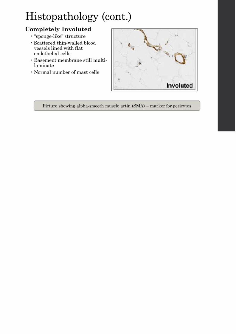
Histopathology (cont.)Completely Involuted
“sponge-like” structure
Scattered thin-walled bloodvessels lined with flatendothelial cells
Basement membrane still multi-laminate
Normal number of mast cells
Picture showing alpha-smooth muscle actin (SMA) – marker for pericyte
8/17/2019 Pedi Hemangioma Pic 2014 03
http://slidepdf.com/reader/full/pedi-hemangioma-pic-2014-03 26/92
Special Locations
C C li i
8/17/2019 Pedi Hemangioma Pic 2014 03
http://slidepdf.com/reader/full/pedi-hemangioma-pic-2014-03 27/92
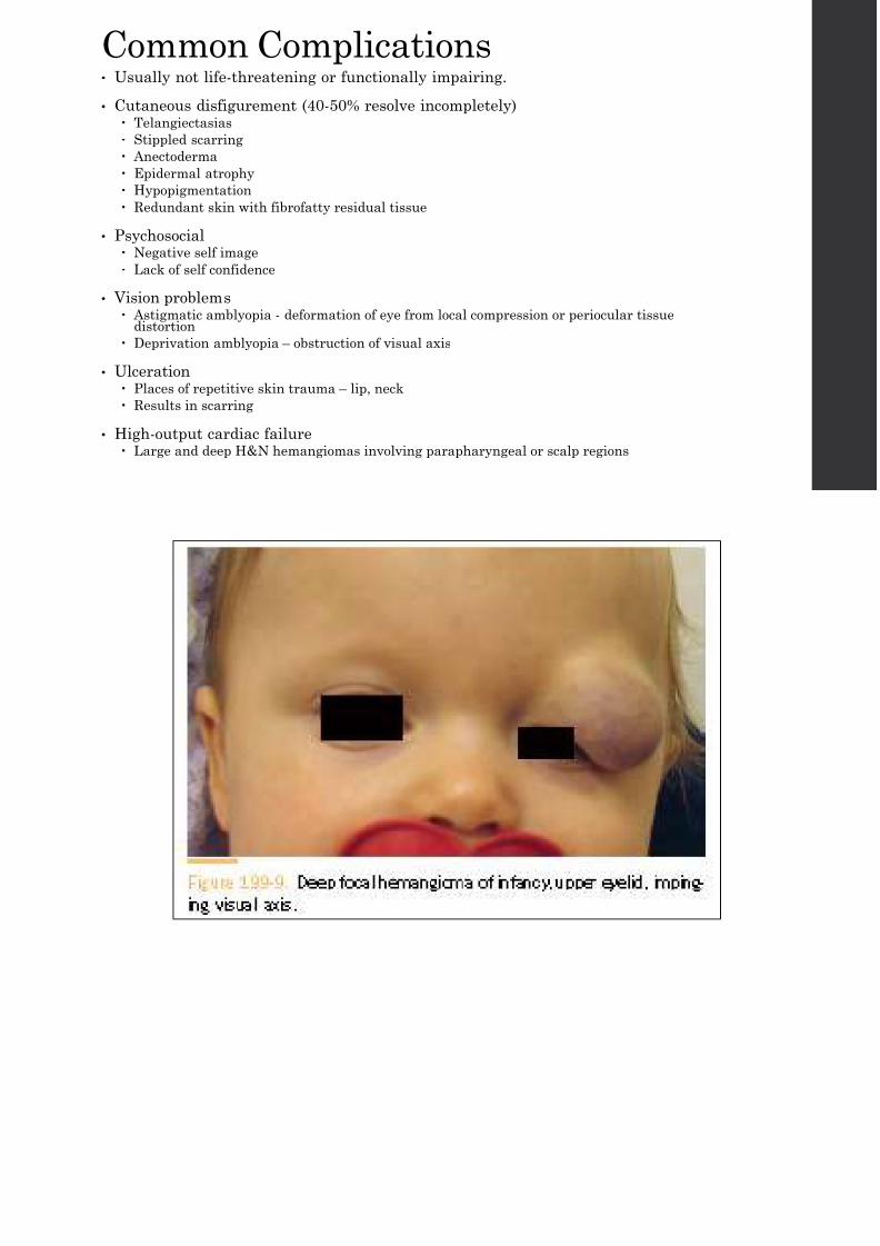
Common Complications• Usually not life-threatening or functionally impairing.
• Cutaneous disfigurement (40-50% resolve incompletely) Telangiectasias
Stippled scarring
Anectoderma Epidermal atrophy
Hypopigmentation
Redundant skin with fibrofatty residual tissue
• Psychosocial Negative self image
Lack of self confidence
• Vision problems
Astigmatic amblyopia - deformation of eye from local compression or periocular tissuedistortion
Deprivation amblyopia – obstruction of visual axis
• Ulceration Places of repetitive skin trauma – lip, neck
Results in scarring
• High-output cardiac failure Large and deep H&N hemangiomas involving parapharyngeal or scalp regions

8/17/2019 Pedi Hemangioma Pic 2014 03
http://slidepdf.com/reader/full/pedi-hemangioma-pic-2014-03 28/92
8/17/2019 Pedi Hemangioma Pic 2014 03
http://slidepdf.com/reader/full/pedi-hemangioma-pic-2014-03 29/92
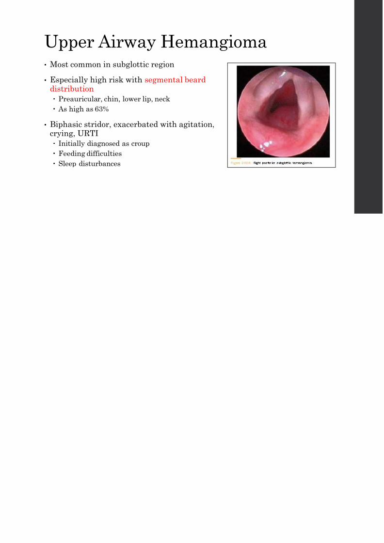
Upper Airway Hemangioma• Most common in subglottic region
• Especially high risk with segmental bearddistribution
Preauricular, chin, lower lip, neck
As high as 63%
• Biphasic stridor, exacerbated with agitation,crying, URTI
Initially diagnosed as croup
Feeding difficulties
Sleep disturbances
8/17/2019 Pedi Hemangioma Pic 2014 03
http://slidepdf.com/reader/full/pedi-hemangioma-pic-2014-03 30/92
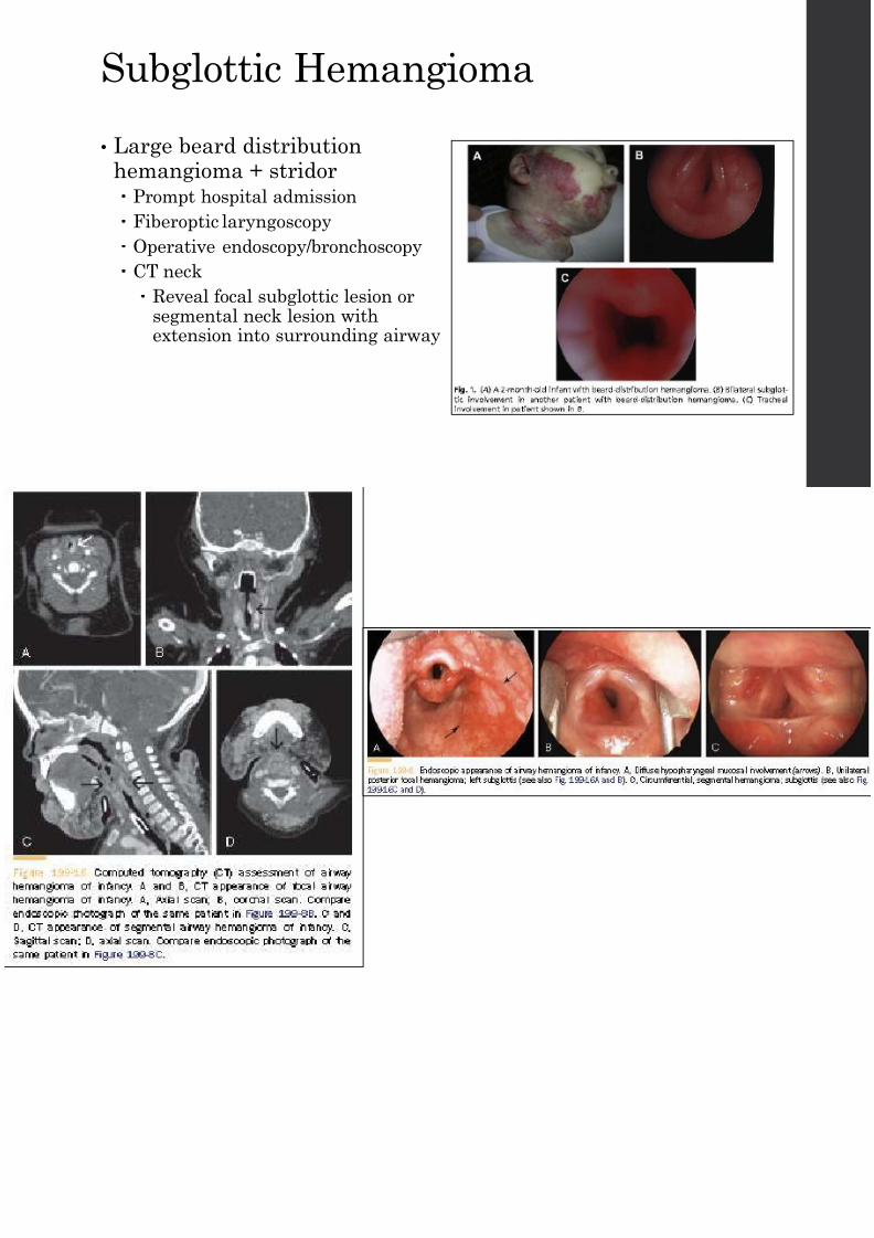
Subglottic Hemangioma
• Large beard distribution
hemangioma + stridor Prompt hospital admission
Fiberoptic laryngoscopy
Operative endoscopy/bronchoscopy
CT neck
Reveal focal subglottic lesion or
segmental neck lesion withextension into surrounding airway
8/17/2019 Pedi Hemangioma Pic 2014 03
http://slidepdf.com/reader/full/pedi-hemangioma-pic-2014-03 31/92
8/17/2019 Pedi Hemangioma Pic 2014 03
http://slidepdf.com/reader/full/pedi-hemangioma-pic-2014-03 32/92
8/17/2019 Pedi Hemangioma Pic 2014 03
http://slidepdf.com/reader/full/pedi-hemangioma-pic-2014-03 33/92
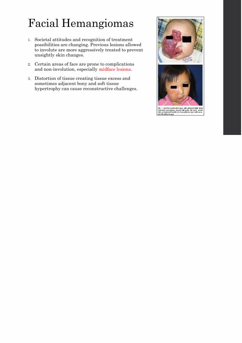
Facial Hemangiomas
1. Societal attitudes and recognition of treatment
possibilities are changing. Previous lesions allowedto involute are more aggressively treated to preventunsightly skin changes.
2. Certain areas of face are prone to complicationsand non-involution, especially midface lesions.
3. Distortion of tissue creating tissue excess andsometimes adjacent bony and soft tissue
hypertrophy can cause reconstructive challenges.
8/17/2019 Pedi Hemangioma Pic 2014 03
http://slidepdf.com/reader/full/pedi-hemangioma-pic-2014-03 34/92
Most common pediatric benign parotid gland tumor
1. Hemangioma
2. Pleomorphic adenoma
3. Mucoepidermoid carcinoma
8/17/2019 Pedi Hemangioma Pic 2014 03
http://slidepdf.com/reader/full/pedi-hemangioma-pic-2014-03 35/92
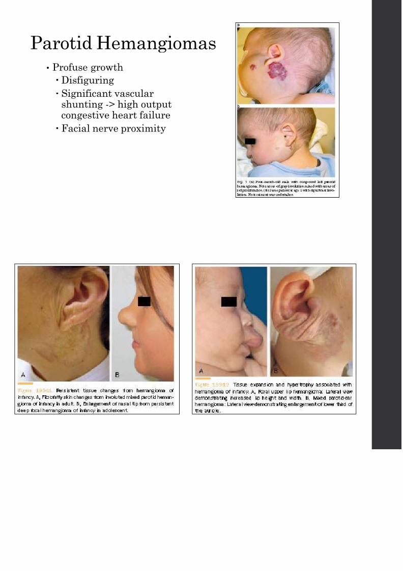
Parotid Hemangiomas
• Profuse growth
Disfiguring
Significant vascularshunting -> high outputcongestive heart failure
Facial nerve proximity
8/17/2019 Pedi Hemangioma Pic 2014 03
http://slidepdf.com/reader/full/pedi-hemangioma-pic-2014-03 36/92
8/17/2019 Pedi Hemangioma Pic 2014 03
http://slidepdf.com/reader/full/pedi-hemangioma-pic-2014-03 37/92
Associated
Syndromes• NOT Kasabach-Merritt Phenomenon
• PHACE Syndrome
8/17/2019 Pedi Hemangioma Pic 2014 03
http://slidepdf.com/reader/full/pedi-hemangioma-pic-2014-03 38/92
Kasabach-Merrit Phenomenon (KM• Described in 1940 by them
• Occurrence of profound thrombocytopenia in association with vasculartumors
KHE and TA, not infantile hemangiomas
• Consult hematology/oncology
• Consumptive coagulopathy
Thrombocytopenia
Hypofibrinogenemia
Elevated D-dimers, PT, aPTT
• Requires extensive chemotherapy for risk of bleeding 2/2 thrombocytopenia
First line: systemic corticosteroids and vincristine
Second line: vincristine, rapamycin, propranolol
Others: cyclophosphamide, aspirin,
Bleeding: platelet transfusion, cryoprecipitate, FFP, aminocaproic acid.
Kaposiform Hemangioendothelioma & Tufted Angiomas
PHACE S d
8/17/2019 Pedi Hemangioma Pic 2014 03
http://slidepdf.com/reader/full/pedi-hemangioma-pic-2014-03 39/92
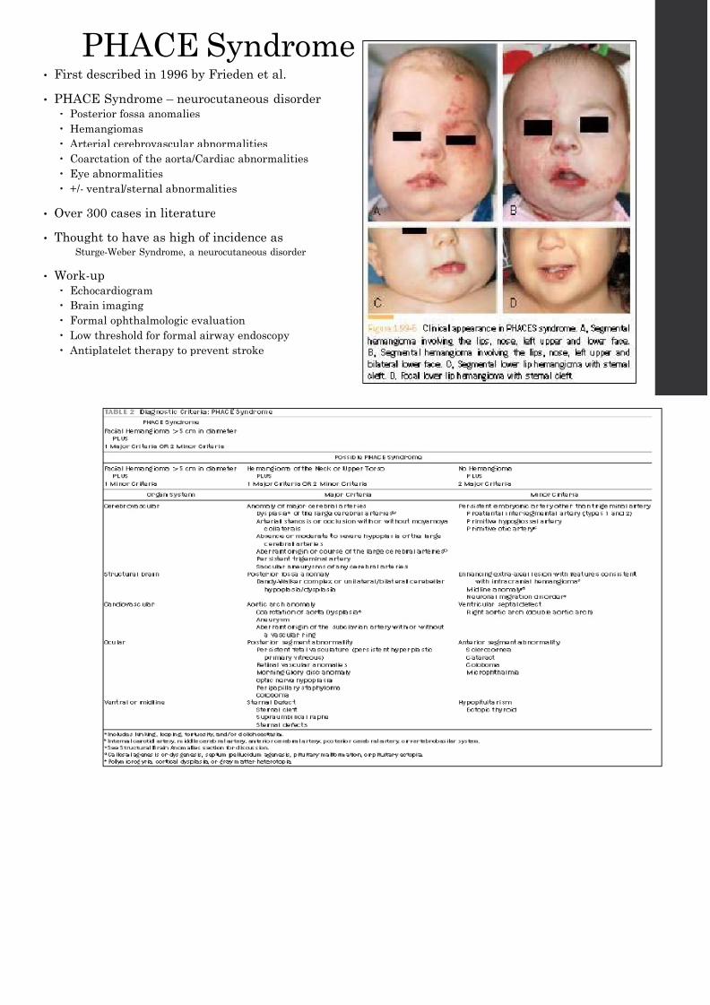
PHACE Syndrome• First described in 1996 by Frieden et al.
• PHACE Syndrome – neurocutaneous disorder
Posterior fossa anomalies
Hemangiomas
Arterial cerebrovascular abnormalities
Coarctation of the aorta/Cardiac abnormalities
Eye abnormalities
+/- ventral/sternal abnormalities
• Over 300 cases in literature
• Thought to have as high of incidence asSturge-Weber Syndrome, a neurocutaneous disorder
• Work-up
Echocardiogram
Brain imaging
Formal ophthalmologic evaluation
Low threshold for formal airway endoscopy
Antiplatelet therapy to prevent stroke

8/17/2019 Pedi Hemangioma Pic 2014 03
http://slidepdf.com/reader/full/pedi-hemangioma-pic-2014-03 40/92
8/17/2019 Pedi Hemangioma Pic 2014 03
http://slidepdf.com/reader/full/pedi-hemangioma-pic-2014-03 41/92
PHACE Syndrome and SGH• Hemangiomas in this syndrome tend to be larger than 5 cm2.
• 98% percent of published PHACE cases report a hemangioma on the face
• Durr et al showed that 52% (12 out of 23) of PHACE syndrome patient wlarge cervicofacial segmental hemangiomas had coexisting subglottichemangiomas.
6 of these 12 were treated at UCSF
5 female, 1 male
3 Caucasian, 3 Hispanic
1 premature at 36 wks, 5 full term
3 had biphasic stridor, 1 had barking cough, 2 without symptoms
Average obstruction of 50%
8/17/2019 Pedi Hemangioma Pic 2014 03
http://slidepdf.com/reader/full/pedi-hemangioma-pic-2014-03 42/92
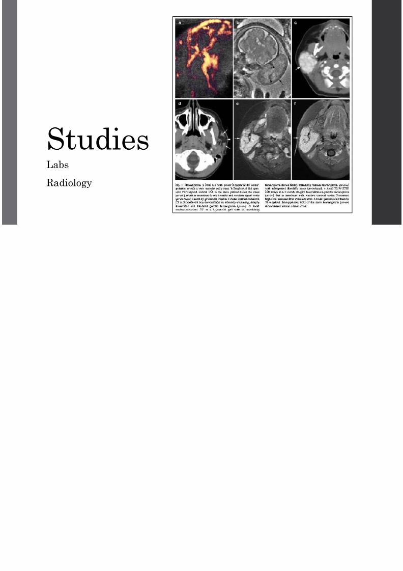
StudiesLabs
Radiology
L b t T t

8/17/2019 Pedi Hemangioma Pic 2014 03
http://slidepdf.com/reader/full/pedi-hemangioma-pic-2014-03 43/92
Laboratory Tests• GLUT-1
• Lewis Antigen (LeY)
• Merosin
• Urinary basic fibroblast growth factor (bFGF) level
Distinguish SGH from vascular or lymphatic malformation
Mast cells produce bFGF
Serial urinary BFGF levels may follow response to any treatment
• Mostly research only
Proliferating cell nuclear antigen
Vascular endothelial growth factor
Type IV collagenase
Urokinase
Radiology
8/17/2019 Pedi Hemangioma Pic 2014 03
http://slidepdf.com/reader/full/pedi-hemangioma-pic-2014-03 44/92
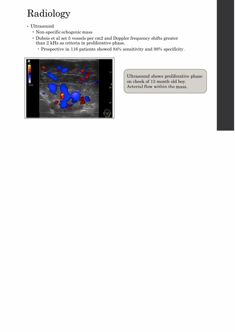
Radiology• Ultrasound
Non-specific echogenic mass
Dubois et al set 5 vessels per cm2 and Doppler frequency shifts greater
than 2 kHz as criteria in proliferative phase. Prospective in 116 patients showed 84% sensitivity and 98% specificity.
Ultrasound shows proliferativ
on cheek of 13 month old boy.
Arterial flow within the mass.
Radiology

8/17/2019 Pedi Hemangioma Pic 2014 03
http://slidepdf.com/reader/full/pedi-hemangioma-pic-2014-03 45/92
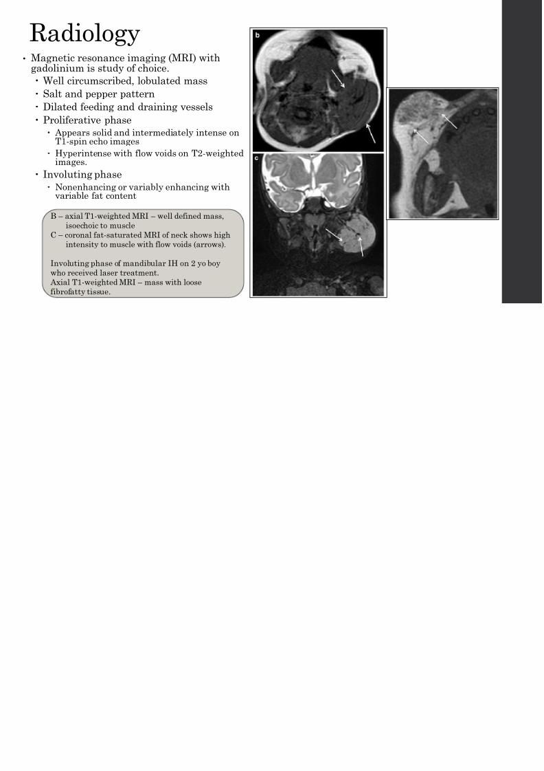
Radiology• Magnetic resonance imaging (MRI) with
gadolinium is study of choice.
Well circumscribed, lobulated mass
Salt and pepper pattern
Dilated feeding and draining vessels Proliferative phase
Appears solid and intermediately intense onT1-spin echo images
Hyperintense with flow voids on T2-weightedimages.
Involuting phase
Nonenhancing or variably enhancing with
variable fat content
B – axial T1-weighted MRI – well defined mass,
isoechoic to muscle
C – coronal fat-saturated MRI of neck shows high
intensity to muscle with flow voids (arrows).
Involuting phase of mandibular IH on 2 yo boy
who received laser treatment.
Axial T1-weighted MRI – mass with loose
fibrofatty tissue.

8/17/2019 Pedi Hemangioma Pic 2014 03
http://slidepdf.com/reader/full/pedi-hemangioma-pic-2014-03 46/92
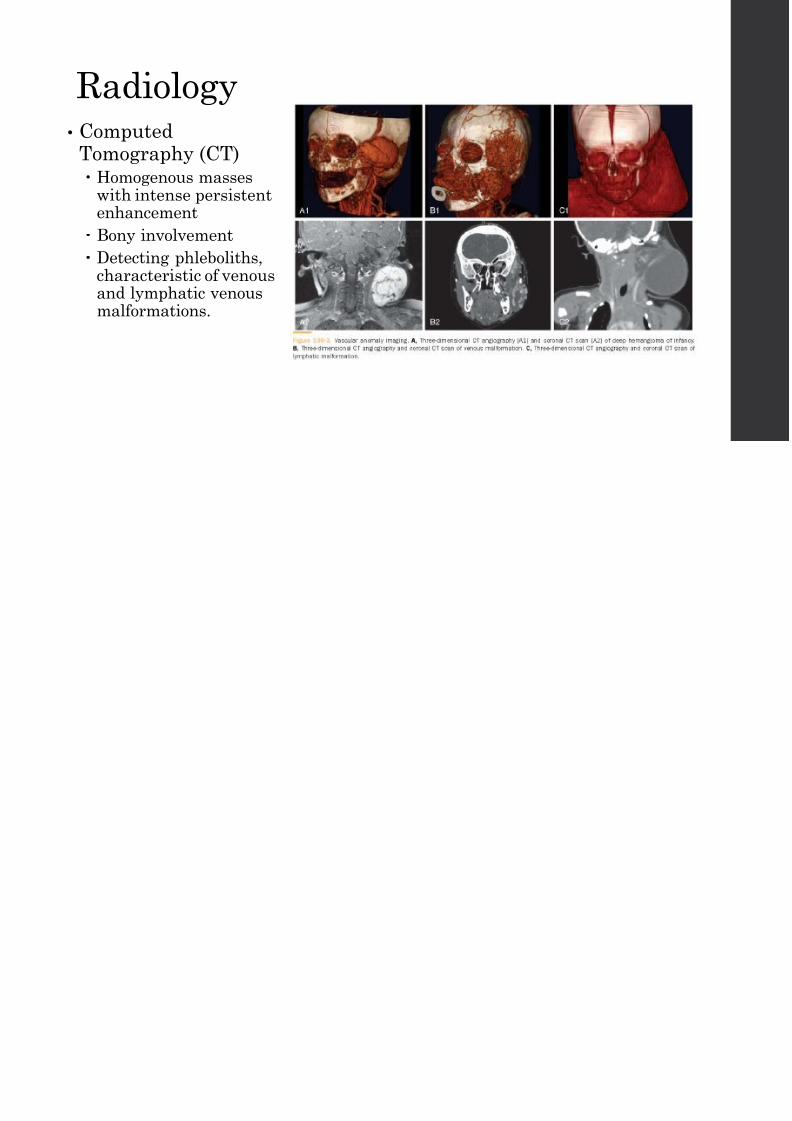
Radiology• Computed
Tomography (CT) Homogenous masses
with intense persistentenhancement
Bony involvement
Detecting phleboliths,characteristic of venous
and lymphatic venousmalformations.
8/17/2019 Pedi Hemangioma Pic 2014 03
http://slidepdf.com/reader/full/pedi-hemangioma-pic-2014-03 47/92
Treatment• No “gold standard”
• Active nonintervention
• Pharmacological
• Surgical
8/17/2019 Pedi Hemangioma Pic 2014 03
http://slidepdf.com/reader/full/pedi-hemangioma-pic-2014-03 48/92
Treatment Principles• Small isolated or multiple skin lesions on the face found soon after
birth should be treated as soon as possible in order to prevent itsprogress into the proliferative phase
• Close observation for involuting hemangiomas.
• Factors:
Size, stage, location, presence of ulceration, cosmetic considerations,functional compromise, psychosocial implications.
Interference with visual axis (eyelid) Risk to cartilage: ear or nose
Airway compromise
Feeding difficulties
W t hf l W iti
8/17/2019 Pedi Hemangioma Pic 2014 03
http://slidepdf.com/reader/full/pedi-hemangioma-pic-2014-03 49/92
Watchful Waiting• Small, stable in non-vital areas
• Observe, record, photography
• Intervention with:
Accelerated growth
Hemorrhage
Infection
Ulceration
Functional problems:
Dysphagia
Trouble breathing
Vision problems
Hearing problems
Leads to high output congestive heart failure
Involves: eyelids, nose, lips, auricle
8/17/2019 Pedi Hemangioma Pic 2014 03
http://slidepdf.com/reader/full/pedi-hemangioma-pic-2014-03 50/92
Pharmacologic Treatment• Corticosteroids
• Interferon alpha-2a
• Imiquimod
• Anti-cancer drugs
Cyclophosphamide
Vincristine
Bleomycin
• Becaplermin, recombinant platelet-derived growth factor, 0.01% gel forulcerations
• Propranolol
8/17/2019 Pedi Hemangioma Pic 2014 03
http://slidepdf.com/reader/full/pedi-hemangioma-pic-2014-03 51/92
Corticosteroids• Historical treatment (>30 yrs) of choice for subglottic and parotid lesions
• Effective during proliferative phase and can slow or cease growth.
• Prednisolone 2 to 5 mg/kg/d for 4-10 wks
• Dose is tapered off
• Up to 90% response rate
• High morbidity
Behavioral changes, hypertension, immunosuppression, gastrointestinalirritation, adrenal insufficiency, cushingoid facies
Inhibition of hypothalamic-pituitary-adrenal axis
• Can be injected intralesionally
Be aware of particle embolization -> blindness
Interferon alpha-2a
8/17/2019 Pedi Hemangioma Pic 2014 03
http://slidepdf.com/reader/full/pedi-hemangioma-pic-2014-03 52/92
Interferon alpha 2a• Anti-angiogenic activity
Can be used in proliferative or involutional stage
• Subcutaneous single daily dose over several months (<3 mo)
• 3 million U/m2
• Monitor CBC, LFT, Coags
• 80-90% response rate
• Common complications Flu-like symptoms, somnolence, anorexia, diarrhea, constipation, neutropenia, elevated
LFTs
• Pertinent complication – epilepsy, spastic diplegia, lower limb disability
Spastic diplegia's particular type of brain damage inhibits the proper development of upper mot
neuron function, impacting the motor cortex, the basal ganglia and the corticospinal tract. Nerv
receptors in the spine leading to affected muscles become unable to properly absorb gamma ami
acid (GABA), the amino acid that regulates muscle tone in humans. Without GABA absorption t
particular nerve rootlets (usually centred, in this case, around the sectors L1-S1 and L2-S2), aff
nerves (here, the ones controlling the legs) perpetually fire the message for their corresponding
permanently, rigidly contract, and the muscles become permanently hypertonic (spastic).
8/17/2019 Pedi Hemangioma Pic 2014 03
http://slidepdf.com/reader/full/pedi-hemangioma-pic-2014-03 53/92
Imiquimod• Imidazole quinolone amine immunomodulatory drug
• Used for herpes, BCC, SCCis, actinic keratosis, lentigo maligna
• Martinez in 2002 attempted to apply this topically in 5% cream every otherday and achieved ideal efficacy.
• Mechanism may be by enhancing immunity through cytokines including:interferon-alpha, IL-6, TNF-alpha
• Sunamura found inhibiting tumor growth and anti-angiogenesis effect of IL-12 may play a role
• Qiu showed a 4% rate of severe local reactions including disfiguringdepigmented scars.
• Mao showed 78.9% of patients with IH had site itching, erythema/edema,peeling, erosion, crusting, ulceration, scarring. 4/19 had fever, nausea ordiarrhea.
8/17/2019 Pedi Hemangioma Pic 2014 03
http://slidepdf.com/reader/full/pedi-hemangioma-pic-2014-03 54/92
Radiation Therapy• Gamma ray produced by radioisotope to bombard nuclei area leading to cell
death.
• 2 Gy per dose for total of 10 Gy maximum
• Radioisotope therapy with strontium-90 used.
• Potential cancer formation

8/17/2019 Pedi Hemangioma Pic 2014 03
http://slidepdf.com/reader/full/pedi-hemangioma-pic-2014-03 55/92
Propranolol: An
incidental discoveryLeaute-Labreze et al discovered Propranolol
2 patient with large segmental cervicofacial hemangiomas were treated witpropranolol when symptoms of high output cardiac failure occurred.
Noted rapid and dramatic regression of the cutaneous lesions as well
The underlying problem in high output failure is a decrease in the systemic
vascular resistance that threatens the arterial blood pressure and causes
activation of neurohormones, resulting in an increase in salt and water
retention by the kidney.
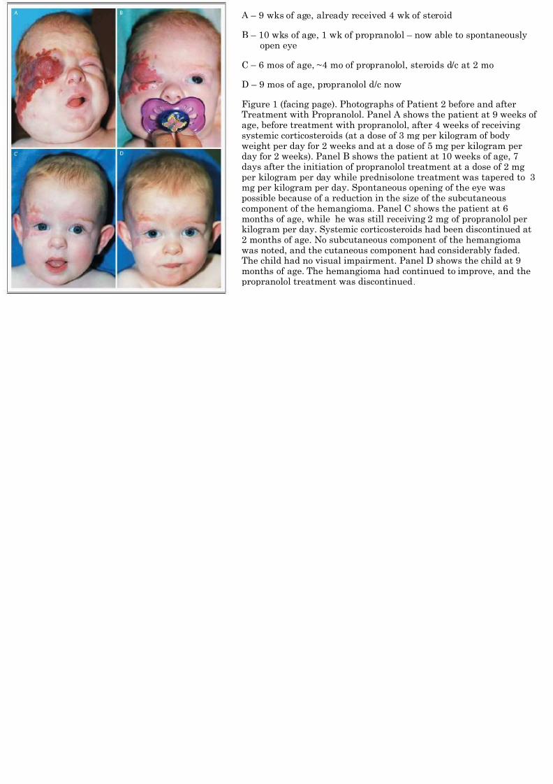
A – 9 wks of age, already received 4 wk of ster
B – 10 wks of age, 1 wk of propranolol – now a
8/17/2019 Pedi Hemangioma Pic 2014 03
http://slidepdf.com/reader/full/pedi-hemangioma-pic-2014-03 56/92
open eye
C – 6 mos of age, ~4 mo of propranolol, steroid
D – 9 mos of age, propranolol d/c now
Figure 1 (facing page). Photographs of Patient Treatment with Propranolol. Panel A shows thage, before treatment with propranolol, after 4systemic corticosteroids (at a dose of 3 mg per kweight per day for 2 weeks and at a dose of 5 mday for 2 weeks). Panel B shows the patient at days after the initiation of propranolol treatmeper kilogram per day while prednisolone treatmmg per kilogram per day. Spontaneous openingpossible because of a reduction in the size of th
component of the hemangioma. Panel C shows months of age, while he was still receiving 2 mkilogram per day. Systemic corticosteroids had2 months of age. No subcutaneous component owas noted, and the cutaneous component had cThe child had no visual impairment. Panel D smonths of age. The hemangioma had continuedpropranolol treatment was discontinued.
Propranolol: Mechanism of Action

8/17/2019 Pedi Hemangioma Pic 2014 03
http://slidepdf.com/reader/full/pedi-hemangioma-pic-2014-03 57/92
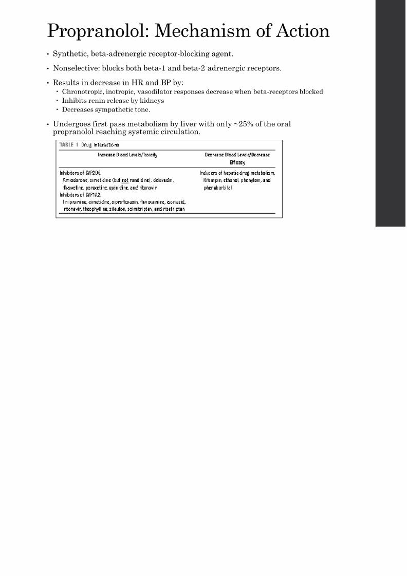
Propranolol: Mechanism of Action• Synthetic, beta-adrenergic receptor-blocking agent.
• Nonselective: blocks both beta-1 and beta-2 adrenergic receptors.
• Results in decrease in HR and BP by: Chronotropic, inotropic, vasodilator responses decrease when beta-receptors blocked
Inhibits renin release by kidneys
Decreases sympathetic tone.
• Undergoes first pass metabolism by liver with only ~25% of the oralpropranolol reaching systemic circulation.
Propranolol
8/17/2019 Pedi Hemangioma Pic 2014 03
http://slidepdf.com/reader/full/pedi-hemangioma-pic-2014-03 58/92
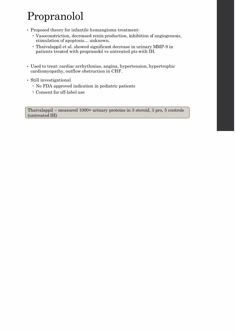
Propranolol• Proposed theory for infantile hemangioma treatment:
Vasoconstriction, decreased renin production, inhibition of angiogenesis,stimulation of apoptosis… unknown.
Thaivalappil et al. showed significant decrease in urinary MMP-9 inpatients treated with propranolol vs untreated pts with IH.
• Used to treat: cardiac arrhythmias, angina, hypertension, hypertrophiccardiomyopathy, outflow obstruction in CHF.
• Still investigational
No FDA approved indication in pediatric patients Consent for off-label use
Thaivalappil – measured 1000+ urinary proteins in 3 steroid, 3 pro, 5 controls
(untreated IH)
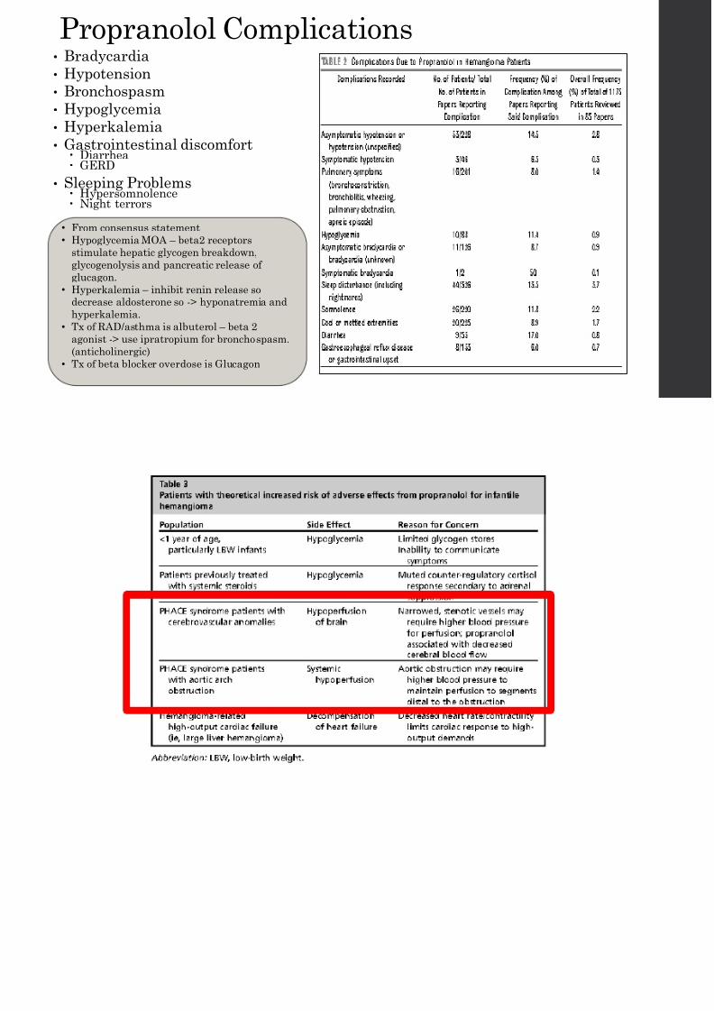
Propranolol Complications• Bradycardia
8/17/2019 Pedi Hemangioma Pic 2014 03
http://slidepdf.com/reader/full/pedi-hemangioma-pic-2014-03 59/92
• Bradycardia
• Hypotension
• Bronchospasm
• Hypoglycemia
• Hyperkalemia
• Gastrointestinal discomfort Diarrhea GERD
• Sleeping Problems Hypersomnolence Night terrors
• From consensus statement
• Hypoglycemia MOA – beta2 receptors
stimulate hepatic glycogen breakdown,
glycogenolysis and pancreatic release ofglucagon.
• Hyperkalemia – inhibit renin release so
decrease aldosterone so -> hyponatremia and
hyperkalemia.
• Tx of RAD/asthma is albuterol – beta 2
agonist -> use ipratropium for bronchospasm.
(anticholinergic)
• Tx of beta blocker overdose is Glucagon
8/17/2019 Pedi Hemangioma Pic 2014 03
http://slidepdf.com/reader/full/pedi-hemangioma-pic-2014-03 60/92
Propranolol Randomized Control Trial
8/17/2019 Pedi Hemangioma Pic 2014 03
http://slidepdf.com/reader/full/pedi-hemangioma-pic-2014-03 61/92
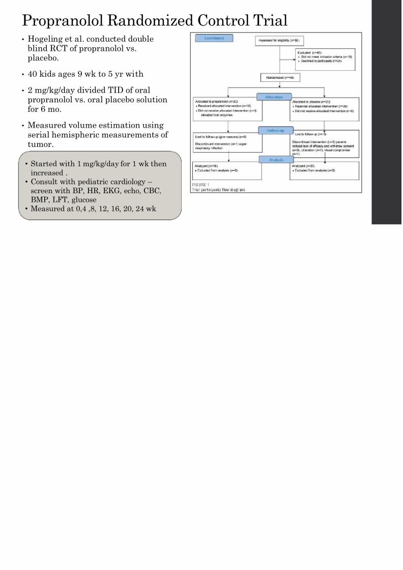
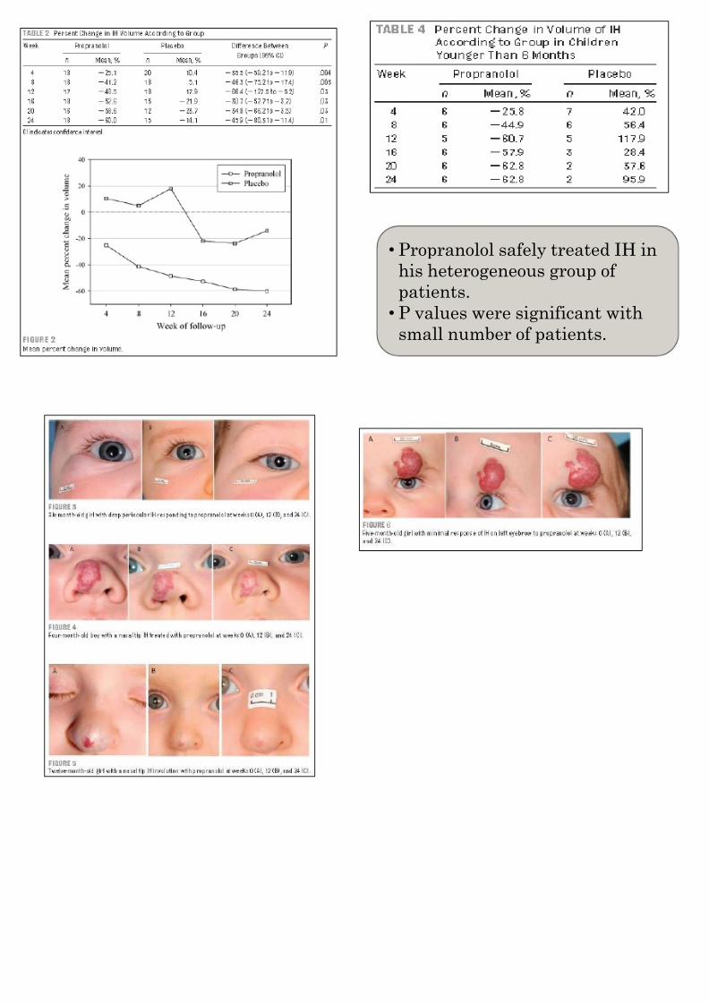
• Hogeling et al. conducted doubleblind RCT of propranolol vs.placebo.
• 40 kids ages 9 wk to 5 yr with
• 2 mg/kg/day divided TID of oralpropranolol vs. oral placebo solutionfor 6 mo.
• Measured volume estimation usingserial hemispheric measurements oftumor.
Color and elevation also assessed
• Started with 1 mg/kg/day for 1 wk then
increased .
• Consult with pediatric cardiology –
screen with BP, HR, EKG, echo, CBC,
BMP, LFT, glucose
• Measured at 0,4 ,8, 12, 16, 20, 24 wk
8/17/2019 Pedi Hemangioma Pic 2014 03
http://slidepdf.com/reader/full/pedi-hemangioma-pic-2014-03 62/92
• Propranolol safely tr
his heterogeneous grpatients.
• P values were signifi
small number of pati
8/17/2019 Pedi Hemangioma Pic 2014 03
http://slidepdf.com/reader/full/pedi-hemangioma-pic-2014-03 63/92

8/17/2019 Pedi Hemangioma Pic 2014 03
http://slidepdf.com/reader/full/pedi-hemangioma-pic-2014-03 64/92
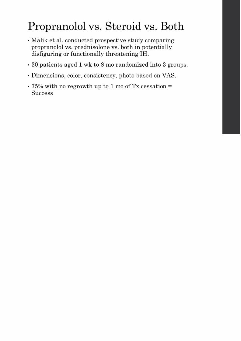
Propranolol vs. Steroid vs. Both• Malik et al. conducted prospective study comparing
propranolol vs. prednisolone vs. both in potentiallydisfiguring or functionally threatening IH.
• 30 patients aged 1 wk to 8 mo randomized into 3 groups.
• Dimensions, color, consistency, photo based on VAS.
• 75% with no regrowth up to 1 mo of Tx cessation =Success
Results
8/17/2019 Pedi Hemangioma Pic 2014 03
http://slidepdf.com/reader/full/pedi-hemangioma-pic-2014-03 65/92
• Mean initial response time (days) [Mean initial response time and consistency chawell defined].
A – 4.1 +/- 3.3 SD
B – 9.78 +/- 7.8 SD
C – 4.7 +/- 3.4 SD
• Consistency change
Very early in A (24 hr) compared to B and C (8 days)
• VAS results [Visual Analog Scale: -10 to +10]
Color fading – A (<48hr) compared to B and C
Flattening – A and C more than B
Mean Reduction in Size – A and C at 3, 6, 12, 18 mo; B only at 6 mo.
• Complications [multiple in a patient with steroids]
A – 2 w/ complications: 1 hypoglycemia, 1 somnolence
B – 9 w/ complications: 5 Cushingoid appearance, 3 GI upset, 3 regrowth during
C – 7 w/ complications: 6 Cushingoid appearance, 4 GI upset, 1 regrowth, 1 infec
Multidisciplinary Strategy
8/17/2019 Pedi Hemangioma Pic 2014 03
http://slidepdf.com/reader/full/pedi-hemangioma-pic-2014-03 66/92
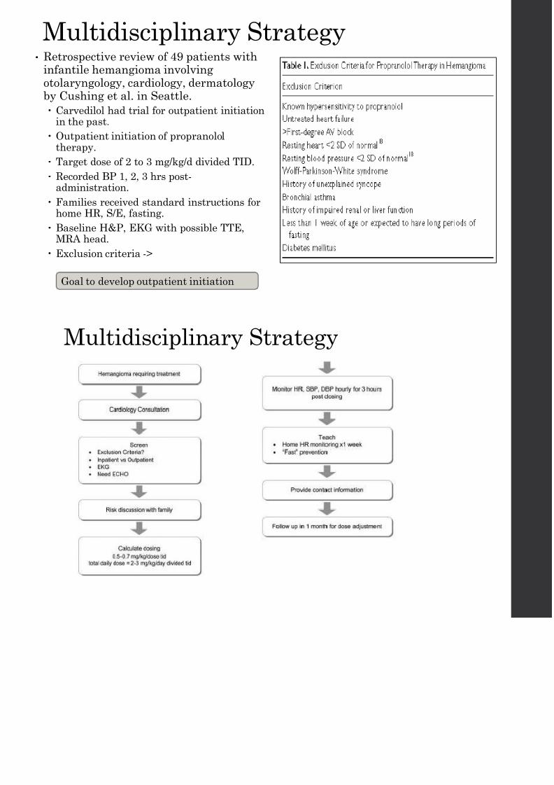
• Retrospective review of 49 patients withinfantile hemangioma involvingotolaryngology, cardiology, dermatologyby Cushing et al. in Seattle.
Carvedilol had trial for outpatient initiationin the past.
Outpatient initiation of propranololtherapy.
Target dose of 2 to 3 mg/kg/d divided TID.
Recorded BP 1, 2, 3 hrs post-administration.
Families received standard instructions forhome HR, S/E, fasting.
Baseline H&P, EKG with possible TTE,MRA head.
Exclusion criteria ->
Goal to develop outpatient initiation
Multidisciplinary Strategy
8/17/2019 Pedi Hemangioma Pic 2014 03
http://slidepdf.com/reader/full/pedi-hemangioma-pic-2014-03 67/92
Multidisciplinary Strategy

8/17/2019 Pedi Hemangioma Pic 2014 03
http://slidepdf.com/reader/full/pedi-hemangioma-pic-2014-03 68/92
• Total of 49 patients
• Mean age at initiation 5.8 +/- 8.4 mo.
• Female:Male 35:9
• 33 involved head and neck, 3 in airway
• 5 patients underwent inpatient therapy
3 had airway compromise, 1 had early heart failure/arrhythmia, 1 social concerns
• HR, SBP, DBP were measured and z scores calculated
HR – no changes in z score greater than 2 from baseline
SBP and DBP – significant changes at only 2 hrs for both absolute and z scores frbaseline
• Conclusion
Outpatient initiation of propranolol for hemangioma is safe with a multidisciplinaapproach with someone experienced with pediatric propranolol therapy.
Propranolol Consensus Statemen
8/17/2019 Pedi Hemangioma Pic 2014 03
http://slidepdf.com/reader/full/pedi-hemangioma-pic-2014-03 69/92
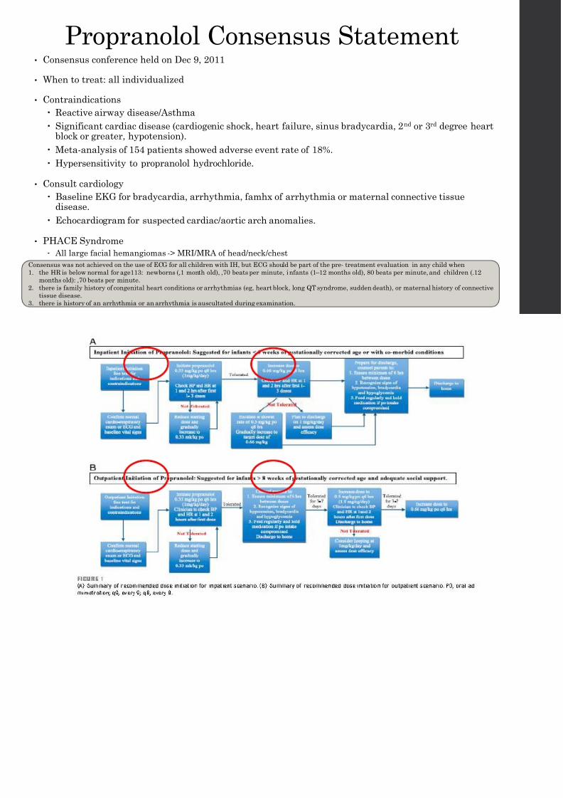
• Consensus conference held on Dec 9, 2011
• When to treat: all individualized
• Contraindications
Reactive airway disease/Asthma Significant cardiac disease (cardiogenic shock, heart failure, sinus bradycardia, 2nd or 3rd de
block or greater, hypotension).
Meta-analysis of 154 patients showed adverse event rate of 18%.
Hypersensitivity to propranolol hydrochloride.
• Consult cardiology
Baseline EKG for bradycardia, arrhythmia, famhx of arrhythmia or maternal connective tisdisease.
Echocardiogram for suspected cardiac/aortic arch anomalies.
• PHACE Syndrome
All large facial hemangiomas -> MRI/MRA of head/neck/chest
Consensus was not achieved on the use of ECG for all children with IH, but ECG should be part of the pre- treatment evaluation in any child
1. the HR is below normal for age113: newborns (,1 month old), ,70 beats per minute, infants (1 – 12 months old), 80 beats per minute, and ch
months old): ,70 beats per minute.
2. there is family history of congenital heart conditions or arrhythmias (eg, heart block, long QT syndrome, sudden death), or maternal histor
tissue disease.
3. there is history of an arrhythmia or an arrhythmia is auscultated during examination.
8/17/2019 Pedi Hemangioma Pic 2014 03
http://slidepdf.com/reader/full/pedi-hemangioma-pic-2014-03 70/92
Propranolol Consensus Statement• Target dose- 2 mg/kg/day divided in 3 doses with a range from 1 and 3 mg/kg/day
8/17/2019 Pedi Hemangioma Pic 2014 03
http://slidepdf.com/reader/full/pedi-hemangioma-pic-2014-03 71/92
• Outpatient Titration - Infants and toddlers older than 8 wks gestation age without signicomorbid conditions.
• Monitor HR & BP
1 set of vitals 1 and 2 hr after initiation or significant dose increase Bradycardia (established criteria)
Newborns (<1 mo old) <70 bpm
Infants (1-12 mo old) <80 bpm
Children (>12 mo old) <70 bpm
Hypotension (systolic BP but not as established)
Newborns (<1 mo old) <57 mmHg (<5th percentile oscillometric) or <64 mmHg (2 SD auscultation)
Infants (1-12 mo old) <85 mmHg (<5th percentile oscillometric) or <65 mmHg (2 SD auscultation)
Children (>12 mo old) <88 mmHg (<5th
percentile oscillometric) or <66 mmHg (2 SD auscultation)
• Make sure to feed at least q4hr
No routine screening for serum glucose recommended.
• Discontinue of concurrent illness, especially in setting or decreased oral intake.
P l l d SGH IH
8/17/2019 Pedi Hemangioma Pic 2014 03
http://slidepdf.com/reader/full/pedi-hemangioma-pic-2014-03 72/92
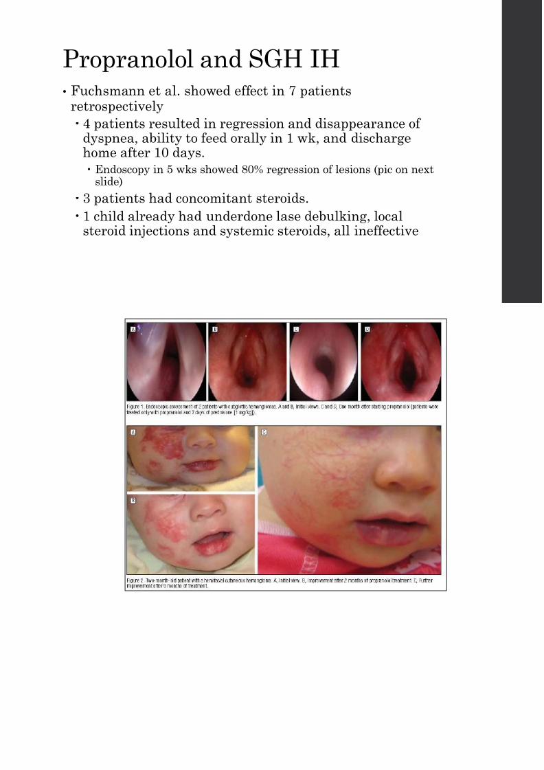
Propranolol and SGH IH• Fuchsmann et al. showed effect in 7 patients
retrospectively 4 patients resulted in regression and disappearance of
dyspnea, ability to feed orally in 1 wk, and dischargehome after 10 days.
Endoscopy in 5 wks showed 80% regression of lesions (pic on nextslide)
3 patients had concomitant steroids.
1 child already had underdone lase debulking, localsteroid injections and systemic steroids, all ineffective

8/17/2019 Pedi Hemangioma Pic 2014 03
http://slidepdf.com/reader/full/pedi-hemangioma-pic-2014-03 73/92
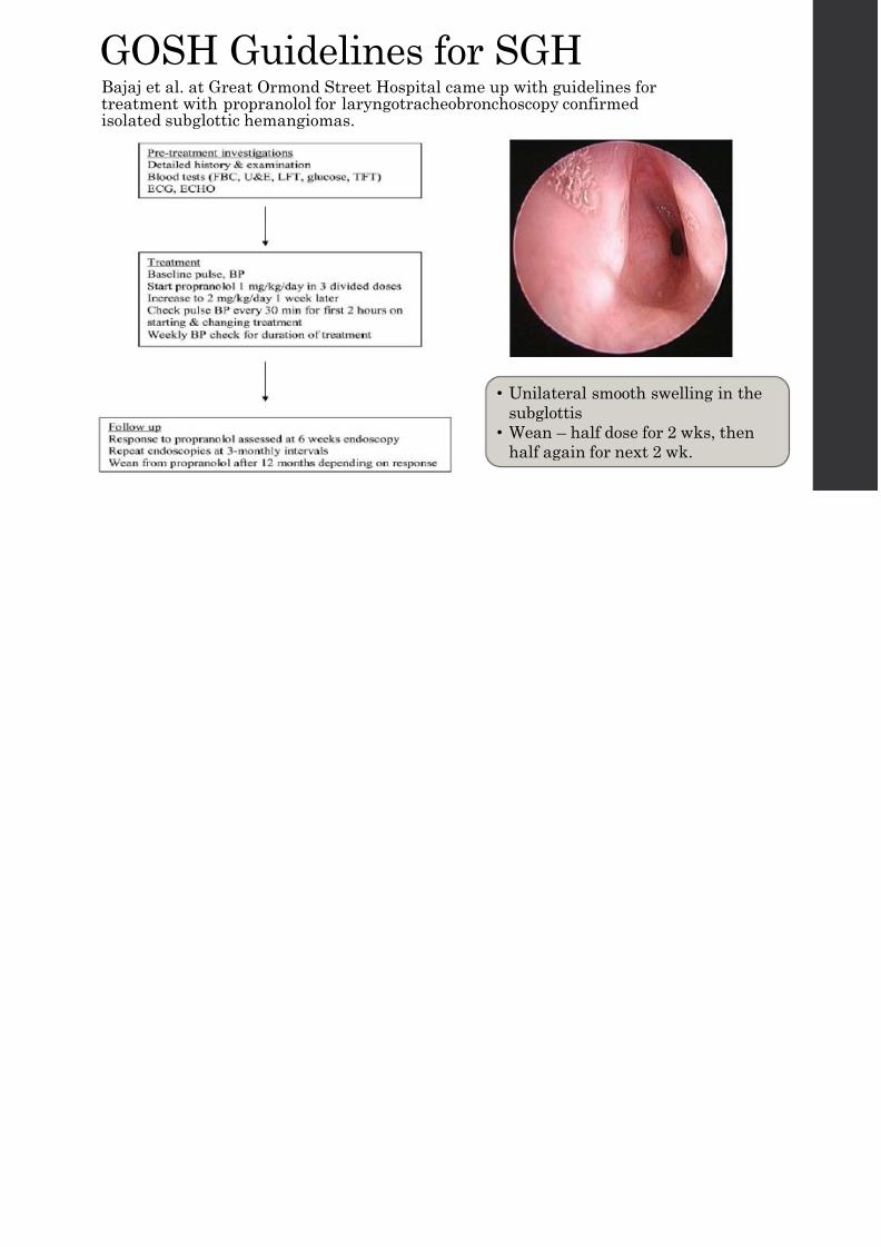
GOSH Guidelines for SGHBajaj et al at Great Ormond Street Hospital came up with guidelines for
8/17/2019 Pedi Hemangioma Pic 2014 03
http://slidepdf.com/reader/full/pedi-hemangioma-pic-2014-03 74/92
Bajaj et al. at Great Ormond Street Hospital came up with guidelines fortreatment with propranolol for laryngotracheobronchoscopy confirmedisolated subglottic hemangiomas.
• Unilateral smooth swellin
subglottis
• Wean – half dose for 2 wk
half again for next 2 wk.
8/17/2019 Pedi Hemangioma Pic 2014 03
http://slidepdf.com/reader/full/pedi-hemangioma-pic-2014-03 75/92
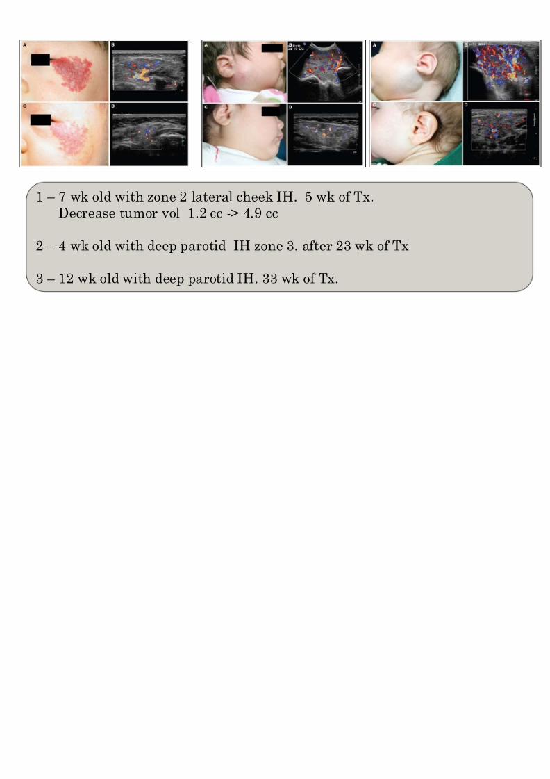
1 – 7 wk old with zone 2 lateral cheek IH. 5 wk of Tx.
Decrease tumor vol 1.2 cc -> 4.9 cc
2 – 4 wk old with deep parotid IH zone 3. after 23 wk of Tx
3 – 12 wk old with deep parotid IH. 33 wk of Tx.

8/17/2019 Pedi Hemangioma Pic 2014 03
http://slidepdf.com/reader/full/pedi-hemangioma-pic-2014-03 76/92
Surgical Options• Tracheostomy
• Cryotherapy
• Laser therapy
• Excision
ABC’s

8/17/2019 Pedi Hemangioma Pic 2014 03
http://slidepdf.com/reader/full/pedi-hemangioma-pic-2014-03 77/92
ABC s
• Airway management for airway IH.
• Tracheostomy to bypass subglottis.
Cryotherapy

8/17/2019 Pedi Hemangioma Pic 2014 03
http://slidepdf.com/reader/full/pedi-hemangioma-pic-2014-03 78/92
• Used in 1960s
• Liquid nitrogen
•
Cellular damage during freezing Intra and extracellular icy crystals form mechanically breaking cellular
membrane.
• Complications:
Cold urticarial
Cryoprecipitate fibrinogen
Cryoglobulinemia
Proliferative or atrophic scarring
Hyperpigmentation or hypopigmentation
Milia
Tissue contracture
Laser Therapy

8/17/2019 Pedi Hemangioma Pic 2014 03
http://slidepdf.com/reader/full/pedi-hemangioma-pic-2014-03 79/92
• Argon Laser
• Pulsed Dye Laser
• Nd:YAG Laser
• KTP Laser
• Indicated for:
Early, superficial hemangiomas
Superficial portion of compound hemangiomas
• Repeated at 2-4 wk intervals
• Choice of laser based on location, size, depth
Argon Laser

8/17/2019 Pedi Hemangioma Pic 2014 03
http://slidepdf.com/reader/full/pedi-hemangioma-pic-2014-03 80/92
Argon Laser• Short wavelength of 488-514 nm
• Unselective thermal damage of blood vessels
• Easy to damage adjacent normal tissue ->scarring/pigmentation
40% treated with Argon Laser may be accompanied byhypertrophic scarring
Limited use
Flash Lamp-pumped Pulsed Dye Laser (FPDL
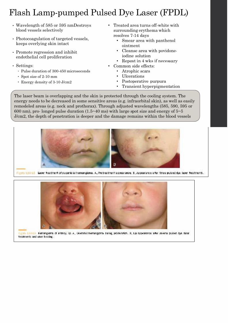
• Wavelength of 585 or 595 nmDestroys • Treated area turns off white with

8/17/2019 Pedi Hemangioma Pic 2014 03
http://slidepdf.com/reader/full/pedi-hemangioma-pic-2014-03 81/92
• Wavelength of 585 or 595 nmDestroysblood vessels selectively
• Photocoagulation of targeted vessels,keeps overlying skin intact
• Promote regression and inhibitendothelial cell proliferation
• Settings:
Pulse duration of 300-450 microseconds
Spot size of 2-10 mm
Energy density of 3-10 J/cm2
• Treated area turns off-white with
surrounding erythema which
resolves 7-14 days
• Smear area with panthenol
ointment• Cleanse area with povidone-
iodine solution
• Repeat in 4 wks if necessary
• Common side effects:
• Atrophic scars
• Ulcerations
• Postoperative purpura
•
Transient hyperpigmentation
The laser beam is overlapping and the skin is protected through the cooling system. T
energy needs to be decreased in some sensitive areas (e.g. infraorbital skin), as well a
remodeled areas (e.g. neck and prothorax). Through adjusted wavelengths (585, 590, 5
600 nm), pro- longed pulse duration (1.5~40 ms) with large spot size and energy of 5~
J/cm2, the depth of penetration is deeper and the damage remains within the blood ve

8/17/2019 Pedi Hemangioma Pic 2014 03
http://slidepdf.com/reader/full/pedi-hemangioma-pic-2014-03 82/92
Nd:YAG

8/17/2019 Pedi Hemangioma Pic 2014 03
http://slidepdf.com/reader/full/pedi-hemangioma-pic-2014-03 83/92
• Neodynium:yttrium aluminum garnet is a solid laser emitcontinuous or pulse type wave of infrared and invisible lig
•
Wavelength of 1064 nm and penetration depth of 4-6 mm• May be utilized for deep hemangiomas
• Very painful so performed under general anesthesia
• 1-4 days after treatment, will remain swollen with possiblblister/scab
• Wound heals 2-4 weeks post-treatment
• Repeated at 5-8 intervals
• Suitable for larger and up to 2 cm deep hemangiomas
KTP Laser

8/17/2019 Pedi Hemangioma Pic 2014 03
http://slidepdf.com/reader/full/pedi-hemangioma-pic-2014-03 84/92
KTP Laser• Potassium titanyl phosphate crystal modifying a 1064 nm
Nd:YAG
• Solid state laser
• Wavelength of 532 nm. Similar to hemoglobin absorption peak
Incidence of postoperative purpura decreases greatly
• Penetration of KTP laser is weaker
• Epidermal melanin also can be targeted leading topigmentation disturbances
CO2 Laser

8/17/2019 Pedi Hemangioma Pic 2014 03
http://slidepdf.com/reader/full/pedi-hemangioma-pic-2014-03 85/92
CO2 Laser
• Removes superficial blood vessels
• Rarely used due to high rate of scar formationand poor effects
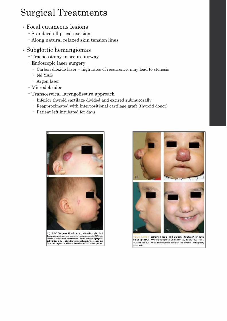
Surgical Treatments
• Focal cutaneous lesions

8/17/2019 Pedi Hemangioma Pic 2014 03
http://slidepdf.com/reader/full/pedi-hemangioma-pic-2014-03 86/92
• Focal cutaneous lesions Standard elliptical excision
Along natural relaxed skin tension lines
• Subglottic hemangiomas Tracheostomy to secure airway
Endoscopic laser surgery
Carbon dioxide laser – high rates of recurrence, may lead to stenosis
Nd:YAG
Argon laser
Microdebrider Transcervical laryngofissure approach
Inferior thyroid cartilage divided and excised submucosally
Reapproximated with interpositional cartilage graft (thyroid donor)
Patient left intubated for days

8/17/2019 Pedi Hemangioma Pic 2014 03
http://slidepdf.com/reader/full/pedi-hemangioma-pic-2014-03 87/92

8/17/2019 Pedi Hemangioma Pic 2014 03
http://slidepdf.com/reader/full/pedi-hemangioma-pic-2014-03 88/92
Conclusion• Infantile hemangiomas are the most common benign tumor in
di i h d d k i l di id

8/17/2019 Pedi Hemangioma Pic 2014 03
http://slidepdf.com/reader/full/pedi-hemangioma-pic-2014-03 89/92
pediatric head and neck, including parotid masses. Distinguish between other vascular tumors and malformations
•
Most do not require active treatment with a predictable prolifeand involution phase.
• When IHs impinge on vital organs or affect aesthetics, treatmebe necessary.
Large segmental head and neck hemangiomas have a highconcurrence of an airway, particularly subglottic, hemangiom
• Propranolol, although mechanisms unknown, has become themainstay treatment for IH over corticosteroids and otherpharmacologic therapies. Requires multi-disciplinary approach for the best individualized managem
the patient.
Board Review

8/17/2019 Pedi Hemangioma Pic 2014 03
http://slidepdf.com/reader/full/pedi-hemangioma-pic-2014-03 90/92
oa d Rev ew
• Infantile hemangiomasGLUT-1 positive
Most common pediatric parotid mass
PHACE Syndrome association
NOT Kasabach-Merritt Phenomenon
Therapy with Propranolol
References• Bajaj Y, et al. Great Ormond Street Hospital treatment guidelines for use of propranolol in infantile isolated subglottic haemangioma. The J of Laryngol
295-8.
• Bingham MM, et al. Propranolol Reduces Infantile Hemangioma Volume and Vessel Density. Otol – Head & Neck Surg 2012; 147: 338-44.

8/17/2019 Pedi Hemangioma Pic 2014 03
http://slidepdf.com/reader/full/pedi-hemangioma-pic-2014-03 91/92
g , p g y g ;
• Celiksoy MH, et al. Management of subglottic hemangioma with propranolol. Amer J of Otol Head and Neck Surg 2014; epub ahead of print.
• Cotton RT, Richardson MA. Advances in head and neck surgery in children. Head and Neck Surgery 1981; 3(5): 424-37.
• Cummings, CW. Otolaryngology, Head and Neck Surgery. Ed. 5. Mosby, 2010.
• Cushing SL, et al. Initial experience with a multidisciplinary strategy for initiation of propranolol therapy for infantile h emangiomas. Otol-Head and Nec78-84.
• Drolet BA, et al. Initiation and use of propranolol for infantile hemangioma: report of a consensus conference. Pediatrics 2012; 128-40.
• Fordham LA, et al. Imaging of congenital vascular and lymphatic anomalies of the head and neck. Neuroimaging Clinic of North America 2000; 10 (1): 11
• Fuchsmann C, et al. Propranolol as First-line Treatment of Head and Neck Hemangiomas. Arch Otolaryngol Head Neck Surg 2011; 137 (5): 471-8.
• Greenberger A, Bischoff J. Pathogenesis of infantile haemangioma. Brit J Dermatol 2013; 169, 12-9.
• Harris GF, White DR. Vascular neoplasms and malformations involving the airway. Facial Plast Surg 2012; 28: 590-5.
• Hartzell LD, Buckmiller LM. Current management of infantile hemangiomas and their associated conditions. Otolaryngology Clinics of North America 20
• Hogeling M, et al. A Randomized Controlled Trial of Propranolol for Infantile Hemangiomas. Pediatrics 2011; 128: e259 -66.
• Holland KE, Drolet BA. Infantile hemangioma. Pediatric Clinic of North America 2010; 57: 1069-83.
• Huoh KC, Rosbe KW. Infantile hemangiomas of the head and neck. Pediatric Clinics of North America 2013; 60: 937-49.
• Leaute-Labreze C, et al. Propranolol for Severe Hemangiomas of Infancy. NEJM 2008; 358: 2649-51.
References (cont.)• Malik AM, et al. Effect of propranolol vs prednisolone vs propranolol with prednisolone in the management of infantile hemangioma: a randomized

8/17/2019 Pedi Hemangioma Pic 2014 03
http://slidepdf.com/reader/full/pedi-hemangioma-pic-2014-03 92/92
Malik AM, et al. Effect of propranolol vs prednisolone vs propranolol with prednisolone in the management of infantile hemangioma: a randomizedcontrolled study. J Pediatric Surg 2013; 48: 2453-59.
• Mao XH, et al. Topical Imiquimod treatment of cutaneous vascular disorders in pediatric patients: clinical evaluation on the efficacy and safety. JZhejiang Univ Sci B; 13 (9): 745-50.
• Metry D, et al. Consensus statement of diagnostic criteria for PHACE Syndrome. Pediatrics 2009; 124: 1447-56.
• Mitchell RB, Pereira KD. Pediatric Otolaryngology for the Clinician. Humana Press, 2009.
• Nozaki T, et al. Imaging of vascular tumors with an emphasis on ISSVA classification. Jpn J Radiol 2013; 31: 775-85.
• Robson CD. Imaging of head and neck neoplasms in children. Pediatric Radiology 2010; 40: 499-509.
• Sie KCY, et al. hemangiomas and vascular malformations of the airway. Otolaryngologic Clinic of North America 2000; 33 (1): 209 -20.
• Thaivalappil S, et al. Propranolol-mediated attenuation of MMP-9 in infants with hemangiomas. JAMA Otol Head Neck Surg 2013; 139 (10): 1026-31.
• Tlougan BE, et al. Medical Management of Tumors Associated With Kasabach-Merritt Phenomenon: An Expert Survey. Pediatric Hematol Oncol2013; 35 (8): 618-22.
• Turkyilmaz Z, et al. Congenital neck masses in children and their embryonic and clinical features. B-ENT 2008; 4: 7-18.
• Virbalas JM, et al. Ped iatric nasal lobular capillary hemangioma. Case Reports in Medicine 2012; epub.
• Yadav P, et a l. Vascular Anomalies of the Head and Neck: A Review of Genetics. Seminars in Ophthalmology 2013; 28 (5 -6): 257-66.
• Zheng JW, et al. A practical guide to treatment of infantile hemangiomas of the head and neck. International Journal of Clinical ExperimentalMedicine 2013; 6 (10): 851-60.