44meera etal
TRANSCRIPT
245
Meera et al., Int J Med Res Health Sci. 2015;4(1):245-247
International Journal of Medical Research
&
Health Scienceswww.ijmrhs.com Volume 4 Issue 1 Coden: IJMRHS Copyright @2014 ISSN: 2319-5886Received: 11
thNov 2014 Revised: 15
thDec 2014 Accepted: 31
stDec 2014
Case report
A LATE ONSET CASE OF SPORADIC DYSCHROMATOSIS UNIVERSALIS HEREDITARIA
*Meera Govindaraju1, Thilak Sundararaj
2, Brindha Thangaraj
3
1Assistant Professor,
2Associate Professor,
3Post graduate student, Department of Dermatology, Meenakshi
Medical College & Research Institute, Kanchipuram
*Corresponding author email: [email protected]
ABSTRACT
Dyschromatosisuniversalishereditaria is an autosomal dominant inherited rare genodermatosis wherein patient
presents with hypopigmented and hyperpigmented macules of varying sizes in a reticulate pattern. We report a
rare case of Dyschromatosisuniversalishereditaria in a 23 year old male patient with no affected family members
suggesting the possibility of sporadic mutation. Patient born of non consanguineous marriage presented with both
hypopigmented macules and hyperpigmentedkeratotic papuleswith progressive diffuse hyperpigmentation over
the trunk and both the extremities. Other system examination was normal. Histopathological examination showed
pigment incontinence with collagenisation of the dermis. A diagnosis of Dyschromatosisuniversalishereditaria
(DUH) was made based on history, clinical morphology and histopathology.
Keywords: Dyschromatosisuniversalis, Reticulate, Genodermatosis, Pigmentation
INTRODUCTION
Dyschromatosisare pigmentarydisorders which
presents with both hyperpigmented and
hypopigmented macules clinically. Various
conditions present with dyschromatosis like
genodermatosis, inflammatory skin diseases,
infections, drugs, chemical use and nutritional
disorders. Two types of dyschromatosis are described
namely dyschromatosisuniversalishereditaria and
dyschromatosissymmetricahereditaria or
acropigmentation of Dohi, wherein there is
predominantly acral distribution as opposed to DUH
with generalized distribution.Autosomal dominant
DUH is a genodermatosis described first by Toyamo1
in Japan in 1929 and again in 1933 in Germany by
Ichikawa and Hiraga. It’s been suggested that DSH
could be a subtype of DUH. Only if, cloning of the
unidentified causative genes is done, we can arrive at
a conclusion whether DSH is a subtype of DUH or
not.
CASEREPORT
A 23 year old unmarried male born of non
consanguineous marriage presented to us with
complaints of diffuse darkening of skin and multiple
black warty skin lesions along with whitish
discolouration over arms, trunk and legs of 5 years
duration. Initially, it started over both the legs and
gradually progressed to involve the thighs and trunk
over last two years. No history of burning or itching
sensation on sun exposure. There was no history
suggestive of any drug intake or chemical exposure.
No other family members suffer from similar skin
lesions.
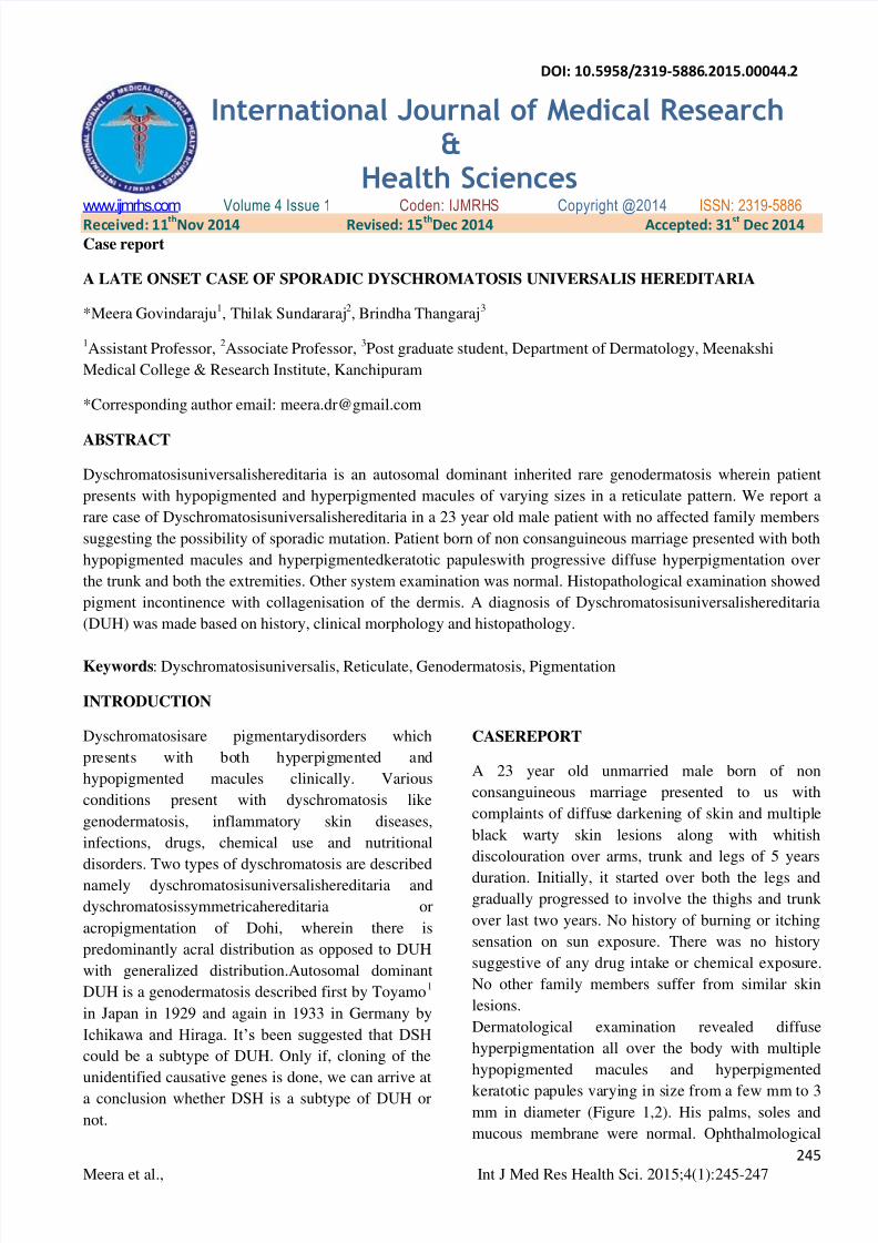
Dermatological examination revealed diffuse
hyperpigmentation all over the body with multiple
hypopigmented macules and hyperpigmented
keratotic papules varying in size from a few mm to 3
mm in diameter (Figure 1,2). His palms, soles and
mucous membrane were normal. Ophthalmological
DOI: 10.5958/2319-5886.2015.00044.2
246
Meera et al., Int J Med Res Health Sci. 2015;4(1):245-247
and ENT examinations were normal. Systemic
examination was normal. All other routine
investigations were within normal limits. VDRL and
HIV were negative. A biopsy was taken from two
sites: hyperpigmentedkeratoticpapule (A) and the
hypopigmented macule (B).
Fig 1: Hyperpigmentedkeratotic papules admixed
with hypopigmented macules over both the legs
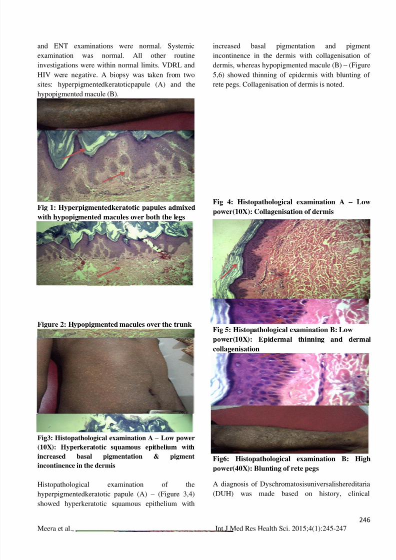
Figure 2: Hypopigmented macules over the trunk
Fig3: Histopathological examination A – Low power
(10X): Hyperkeratotic squamous epithelium with
increased basal pigmentation & pigment
incontinence in the dermis
Histopathological examination of the
hyperpigmentedkeratotic papule (A) – (Figure 3,4)
showed hyperkeratotic squamous epithelium with
increased basal pigmentation and pigment
incontinence in the dermis with collagenisation of
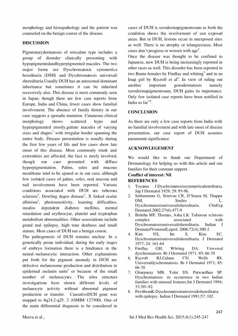
dermis, whereas hypopigmented macule (B) – (Figure
5,6) showed thinning of epidermis with blunting of
rete pegs. Collagenisation of dermis is noted.
Fig 4: Histopathological examination A – Low
power(10X): Collagenisation of dermis
Fig 5: Histopathological examination B: Low
power(10X): Epidermal thinning and dermal
collagenisation
Fig6: Histopathological examination B: High
power(40X): Blunting of rete pegs
A diagnosis of Dyschromatosisuniversalishereditaria
(DUH) was made based on history, clinical
247
Meera et al., Int J Med Res Health Sci. 2015;4(1):245-247
morphology and histopathology and the patient was
counseled on the benign course of the disease.
DISCUSSION
Pigmentarydermatosis of reticulate type includes a
group of disorder clinically presenting with
hypopigmentedandhyperpigmented macules. The two
major forms are Dyschromatosis symmetrica
hereditaria (DSH) and Dyschromatosis universali
shereditaria.Usually DUH has an autosomal dominant
inheritance but sometimes it can be inherited
recessively also. This disease is most commonly seen
in Japan; though there are few case reports from
Europe, India and China, fewer cases show familial
involvement. The absence of family history in our
case suggests a sporadic mutation. Cutaneous clinical
morphology shows scattered hypo and
hyperpigmented mostly,guttate macules of varying
sizes and shapes2
with irregular border spanning the
entire body. Disease presentation is usually during
the first few years of life and few cases show late
onset of this disease. Most commonly trunk and
extremities are affected, the face is rarely involved,
though our case presented with diffuse
hyperpigmentation. Palms, soles and mucous
membrane tend to be spared as in our case, although
few isolated cases of palms, soles, oral mucosa and
nail involvement have been reported. Various
conditions associated with DUH are tuberous
sclerosis3, Dowling degos disease
3, X linked ocular
albinism3, photosensitivity, learning difficulties,
insulin- dependent diabetes mellitus, mental
retardation and erythrocyte, platelet and tryptophan
metabolism abnormalities. Other associations include
grand mal epilepsy, high tone deafness and small
stature. Most cases of DUH run a benign course.
The pathogenesis of DUH remains unclear. In a
genetically prone individual, during the early stages
of embryo formation there is a hindrance in the
neural – melanocytic interaction. Other explanations
put forth for the pigment anomaly in DUH are
defective melanosome production and distribution in
epidermal melanin units3
or because of the small
number of melanocytes. The ultra structure
investigations have shown different levels of
melanocyte activity without abnormal pigment
production or transfer. CausativeDUH gene was
mapped to 6q24.2-q25. 2 (OMIM 127500). One of
the main differential diagnosis to be considered in
cases of DUH is xerodermapigmentosum as both the
condition shows the involvement of sun exposed
areas. But in DUH, lesions occur in unexposed sites
as well. There is no atrophy or telangiectasia. Most
cases don’t progress or worsen with age4.
Once the disease was thought to be confined to
Japanese, now DUH is being increasingly reported in
other races as well. This disorder has been reported in
two Bantu females by Findlay and whiting5
and in an
Iraqi girl by Rycroft et al6. In view of ruling out
another important genodermatosis namely
xerodermapigmentosum, DUH gains its importance.
Only few isolated case reports have been notified in
India so far7,8
.
CONCLUSION
As there are only a few case reports from India with
no familial involvement and with late onset of disease
presentation, our case report of DUH assumes
paramount significance
ACKNOWLEGEMENT
We would like to thank our Department of
Dermatology for helping us with this article and our
families for their constant support.
Conflict of interest: Nil
REFERENCES
1. Toyama J.Dyschromatosissymmetricahereditaria.
Jap J Dermatol 1929; 29: 95-96.2. Sethuraman G, Srinivas CR, D’Souza M, Thappa
DM, Smiles L.Dyschromatosisuniversalishereditaria. ClinExp
Dermatol.2002;27(6):477-9
3. Binitha MP, Thomas, Asha LK. Tuberous sclerosiscomplex associated with
Dyschromatosisuniversalishereditaria. Indian JDermatolVenereolLeprol. 2006;72(4);300-2
4. Kim NS, Im S, Kim SC.
Dyschromatosisuniversalishereditaria. J Dermatol1977; 24: 161-64
5. Findlay GH, Whiting DA. Universal
dyschromatosis. Br J Dermatol 1971; 85; 66-706. Rycroft RJ,Calnan CD, Wells RS.
Universaldyschromatosis. Br J Dermatol 1971; 85:66-70
7. Gharpuray MB, Tolat SN, Patwardhan SP.
Dyschromatosis: its occurrence in two Indianfamilies with unusual features.Int J Dermatol 1994;
33:391-92.8. PavithranK.Dyschromatosisuniversalishereditaria
with epilepsy. Indian J Dermatol 1991;57: 102