52 rohit etal
TRANSCRIPT
8/20/2019 52 Rohit Etal
http://slidepdf.com/reader/full/52-rohit-etal 1/3
737
Rohit et al., Int J Med Res Health Sci. 2015;4(3):737-739
International Journal of Medical Research
&
Health Scienceswww.ijmrhs.com Volume 4 Issue 3 Coden: IJMRHS Copyright @2015 ISSN: 2319-5886Received: 29th May 2015 Revised: 15th June 2015 Accepted: 22nd June 2015
Case report
TYPHOID AFTERMATH: PRESENTING AS VASCULITIS, NEURORETINITIS AND MACULAR
NEUROSENSORY DETACHMENT
*Rohit Laul1, Atif Ali MIR
2, Shazia Shafi
3
1MS FERC,
2MS FVRS, DNB MNAMS, Practitioners, Dr. Agarwal's Eye Hospital, Chennai, Tamil Nadu, India
*Corresponding author email: [email protected]
ABSTRACT
Systemic immune-mediated reactions are known to occur following typhoid illness. Vasculitis, neuroretinitis and
macular neurosensory detachment are amongst the rarely documented aftermath of typhoid fever. A 32-year-old
male came with complaints of decreased vision in both eyes with history of typhoid fever (treated adequately 4
weeks prior and declared cured 2 weeks prior to ocular manifestations) who was found to have vasculitis,
neuroretinitis and neurosensory detachment at macula. His BCVA was 1/60 in right eye and 6/12 in left eye. The
inflammation completely resolved and there was marked improvement in the visual acuity, 6/12 in right eye and
6/6 in left eye after treatment with oral steroids. Immune-mediated vasculitis and neuroretinitis, the ocular
aftermath of typhoid fever responded well to oral steroids
Keywords: Neuroretinitis, Immune-mediated neuroretinitis
INTRODUCTION
Salmonella typhi causes typhoid infection. It can give
rise to fever, gastroenteritis and
septicemia. Salmonella rarely affects ocular tissue[1]
.
This may be direct infection or due to an immune-
mediated mechanism. Ocular complications of
typhoid fever include iritis, choroiditis, retinal
hemorrhage, panophthalmitis and endophthalmitis as
reported by Hersing and Duke-Elders. After complete
resolution of typhoid fever, there are documented
evidences of endophthalmitis[2,3]
. Here we are
reporting a patient who presented with
neuroretinitis and had typhoid fever 4weeks prior to
presentation
CASE REPORT
A 32-year-old Indian male presented to Dr Agarwal's
Eye Hospital, Salem with sudden, painless diminution
of vision in both the eyes (right eye > left eye) for 2
weeks. He gave a past history of typhoid fever 4
weeks ago. Lab investigations during the fever
showed positive Widal test with high `O' antigen
(1:160) and `H' antigen (1:40) titres, while `AH' and
`BH' antigens were non-reactive. At the onset fever,
treatment was initiated with oral ofloxacin 400 mg
twice daily for 14 days; following which, the fever
resolved. He started experiencing diminution of
vision 2 weeks after the completion of treatment. At
presentation, his best corrected visual acuity was
1/60,in the right eye and 6/12 in the left eye. On
examination with slit lamp, anterior segments of both
eyes were within normal limits except for relative
afferent pupillary defect in the right eye. Colour
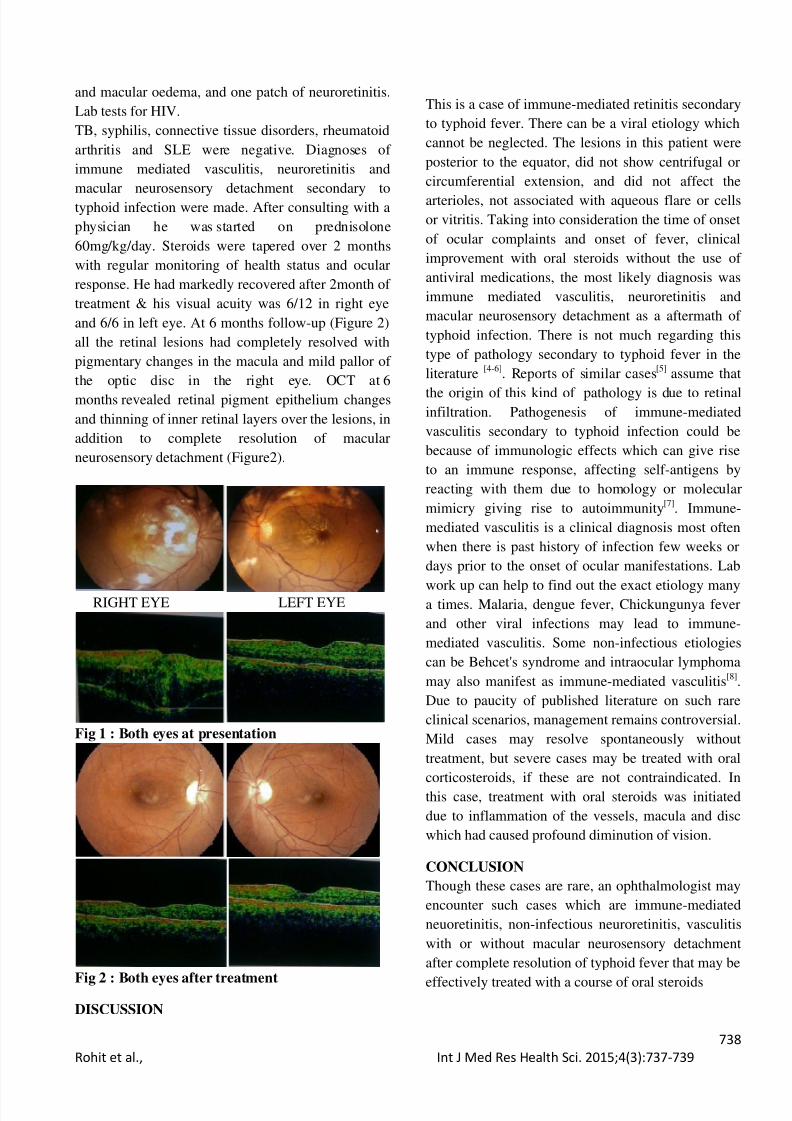
vision was defective in right eye. On fundus
examination media was clear, mild disc pallor was
noted. Vasculitis with multiple areas of deep
neuroretinitis was seen. Macular neurosensory
detachment was evident on OCT scans (Figure 1).The left eye (Figure 1) had clear media, normal disc
DOI: 10 5958/2319 5886 2015 00143 5
8/20/2019 52 Rohit Etal
http://slidepdf.com/reader/full/52-rohit-etal 2/3
738
Rohit et al., Int J Med Res Health Sci. 2015;4(3):737-739
and macular oedema, and one patch of neuroretinitis.
Lab tests for HIV.
TB, syphilis, connective tissue disorders, rheumatoid
arthritis and SLE were negative. Diagnoses of
immune mediated vasculitis, neuroretinitis and
macular neurosensory detachment secondary to
typhoid infection were made. After consulting with a
physician he was started on prednisolone
60mg/kg/day. Steroids were tapered over 2 months
with regular monitoring of health status and ocular
response. He had markedly recovered after 2month of
treatment & his visual acuity was 6/12 in right eye
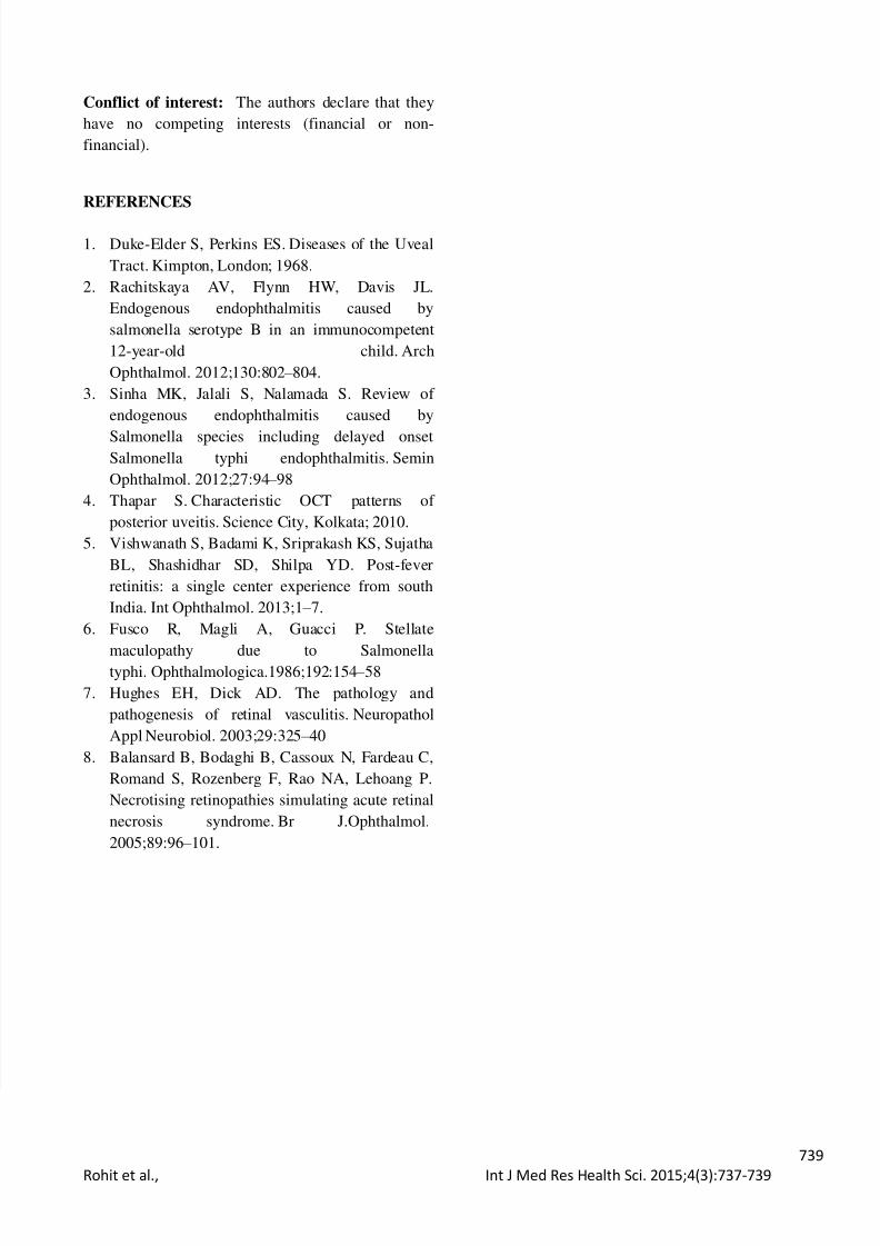
and 6/6 in left eye. At 6 months follow-up (Figure 2)
all the retinal lesions had completely resolved with
pigmentary changes in the macula and mild pallor of
the optic disc in the right eye. OCT at 6
months revealed retinal pigment epithelium changes
and thinning of inner retinal layers over the lesions, in
addition to complete resolution of macular
neurosensory detachment (Figure2).
RIGHT EYE LEFT EYE
Fig 1 : Both eyes at presentation
Fig 2 : Both eyes after treatment
DISCUSSION
This is a case of immune-mediated retinitis secondary
to typhoid fever. There can be a viral etiology which
cannot be neglected. The lesions in this patient were
posterior to the equator, did not show centrifugal or
circumferential extension, and did not affect the
arterioles, not associated with aqueous flare or cells
or vitritis. Taking into consideration the time of onset
of ocular complaints and onset of fever, clinical
improvement with oral steroids without the use of
antiviral medications, the most likely diagnosis was
immune mediated vasculitis, neuroretinitis and
macular neurosensory detachment as a aftermath of
typhoid infection. There is not much regarding this
type of pathology secondary to typhoid fever in the
literature[4-6]
. Reports of similar cases[5]
assume that
the origin of this kind of pathology is due to retinalinfiltration. Pathogenesis of immune-mediated
vasculitis secondary to typhoid infection could be
because of immunologic effects which can give rise
to an immune response, affecting self-antigens by
reacting with them due to homology or molecular
mimicry giving rise to autoimmunity[7]
. Immune-
mediated vasculitis is a clinical diagnosis most often
when there is past history of infection few weeks or
days prior to the onset of ocular manifestations. Lab
work up can help to find out the exact etiology manya times. Malaria, dengue fever, Chickungunya fever
and other viral infections may lead to immune-
mediated vasculitis. Some non-infectious etiologies
can be Behcet's syndrome and intraocular lymphoma
may also manifest as immune-mediated vasculitis[8]
.
Due to paucity of published literature on such rare
clinical scenarios, management remains controversial.
Mild cases may resolve spontaneously without
treatment, but severe cases may be treated with oral
corticosteroids, if these are not contraindicated. In
this case, treatment with oral steroids was initiated
due to inflammation of the vessels, macula and disc
which had caused profound diminution of vision.
CONCLUSION
Though these cases are rare, an ophthalmologist may
encounter such cases which are immune-mediated
neuoretinitis, non-infectious neuroretinitis, vasculitis
with or without macular neurosensory detachment
after complete resolution of typhoid fever that may be
effectively treated with a course of oral steroids
8/20/2019 52 Rohit Etal
http://slidepdf.com/reader/full/52-rohit-etal 3/3
739
Rohit et al., Int J Med Res Health Sci. 2015;4(3):737-739
Conflict of interest: The authors declare that they
have no competing interests (financial or non-
financial).
REFERENCES
1. Duke-Elder S, Perkins ES. Diseases of the Uveal
Tract. Kimpton, London; 1968.
2. Rachitskaya AV, Flynn HW, Davis JL.
Endogenous endophthalmitis caused by
salmonella serotype B in an immunocompetent
12-year-old child. Arch
Ophthalmol. 2012;130:802 – 804.
3. Sinha MK, Jalali S, Nalamada S. Review of
endogenous endophthalmitis caused by
Salmonella species including delayed onsetSalmonella typhi endophthalmitis. Semin
Ophthalmol. 2012;27:94 – 98
4. Thapar S. Characteristic OCT patterns of
posterior uveitis. Science City, Kolkata; 2010.
5. Vishwanath S, Badami K, Sriprakash KS, Sujatha
BL, Shashidhar SD, Shilpa YD. Post-fever
retinitis: a single center experience from south
India. Int Ophthalmol. 2013;1 – 7.
6. Fusco R, Magli A, Guacci P. Stellate
maculopathy due to Salmonellatyphi. Ophthalmologica.1986;192:154 – 58
7. Hughes EH, Dick AD. The pathology and
pathogenesis of retinal vasculitis. Neuropathol
Appl Neurobiol. 2003;29:325 – 40
8. Balansard B, Bodaghi B, Cassoux N, Fardeau C,
Romand S, Rozenberg F, Rao NA, Lehoang P.
Necrotising retinopathies simulating acute retinal
necrosis syndrome. Br J.Ophthalmol.
2005;89:96 – 101.